



Investigation into the competitive quenching mechanism of Cu2+ and Fe3+ on nitrogen-doped carbon dots based on a four-state kinetic model
-
摘要:
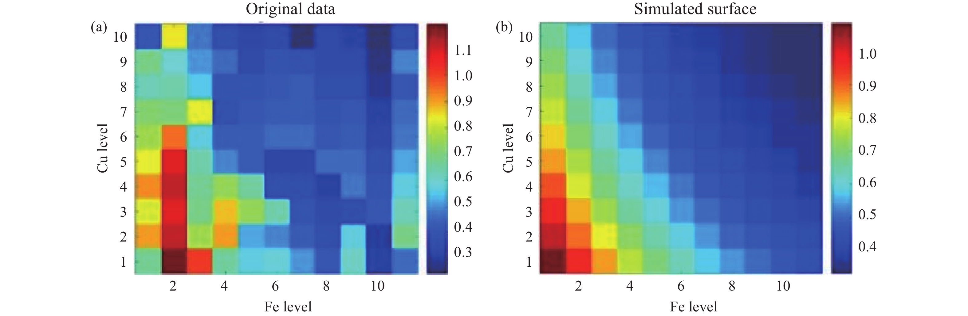
由于实际水环境中多离子共存时的原位拮抗与位点竞争导致传统线性传感模型失效,本文以一步水热法合成的氮掺杂碳点(N-CDs)为平台,通过构建二维交叉荧光响应矩阵,研究了Cu2+与Fe3+共存体系下的微观响应机制。首先,实验观测了不同干扰背景下的荧光猝灭演化规律。结果显示,在高浓度Cu2+背景下,Fe3+诱导的荧光响应表现出显著的非线性偏移与猝灭阻滞,证实两种离子在纳米界面存在激烈的排他性抢夺。随后,为解析该非线性过程,基于细致平衡原理构建了“四态物理动力学模型”,并推导出内含热力学协同因子(
$ \alpha $ )的全局响应解析式。最后,利用理论模型对实验响应矩阵进行了全局曲面拟合。结果表明,理论拟合与实验数据高度吻合,提取出协同因子$ \alpha $ ≈ 0.015。该极低数值定量证实了由高价态离子构筑的极端物理屏蔽与静电排斥效应。本研究将交叉干扰转化为可量化的本征热力学参数,为复杂体系非线性信号解码及界面动力学研究提供了坚实的理论基础。Abstract:Due to the in-situ antagonism and site competition during multi-ion coexistence in real water environments, traditional linear sensing models often fail. In this study, nitrogen-doped carbon dots (N-CDs) synthesized via a one-step hydrothermal method were used as a platform to investigate the microscopic response mechanism in Cu2+ and Fe3+ coexisting systems by constructing a 2D cross-fluorescence response matrix. Firstly, the fluorescence quenching evolution under different interference backgrounds was experimentally observed. The results showed that under a high concentration Cu2+ background, the fluorescence response induced by Fe3+ exhibited significant nonlinear shifts and quenching stagnation, confirming the intense exclusive competition between the two ions at the nano-interface. Subsequently, to analyze this nonlinear process, a "four-state physical kinetic model" was constructed based on the principle of detailed balance, and a global response analytical expression containing the thermodynamic synergy factor (
$ \alpha $ ) was derived. Finally, a global surface fitting was performed on the experimental response matrix using the theoretical model. The results demonstrated a high degree of agreement between the theoretical fit and experimental data, yielding a synergy factor of$ \alpha $ ≈ 0.015. This extremely low value quantitatively confirms the extreme physical shielding and electrostatic repulsion effects constructed by high-valent ions. This study transforms cross-interference into quantifiable intrinsic thermodynamic parameters providing a solid theoretical foundation for nonlinear signal decoding and interface kinetics research in complex systems. -
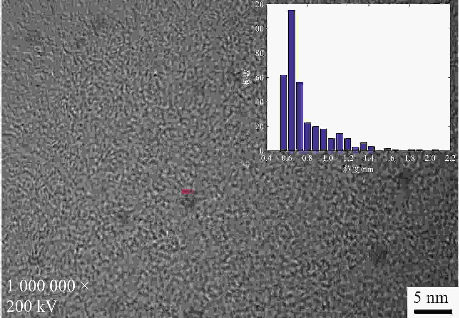
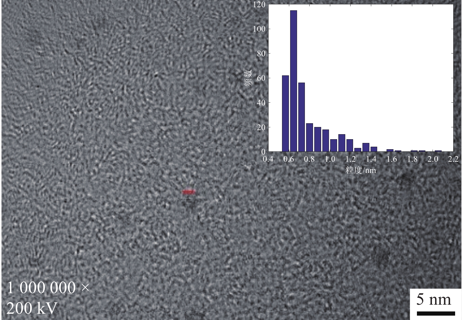
图 1 碳点的TEM图片, 插图为粒径分布直方图
Figure 1. Transmission electron microscopy (TEM) image of the nitrogen-doped carbon dots (N-CDs). The inset shows the corresponding particle size distribution histogram.
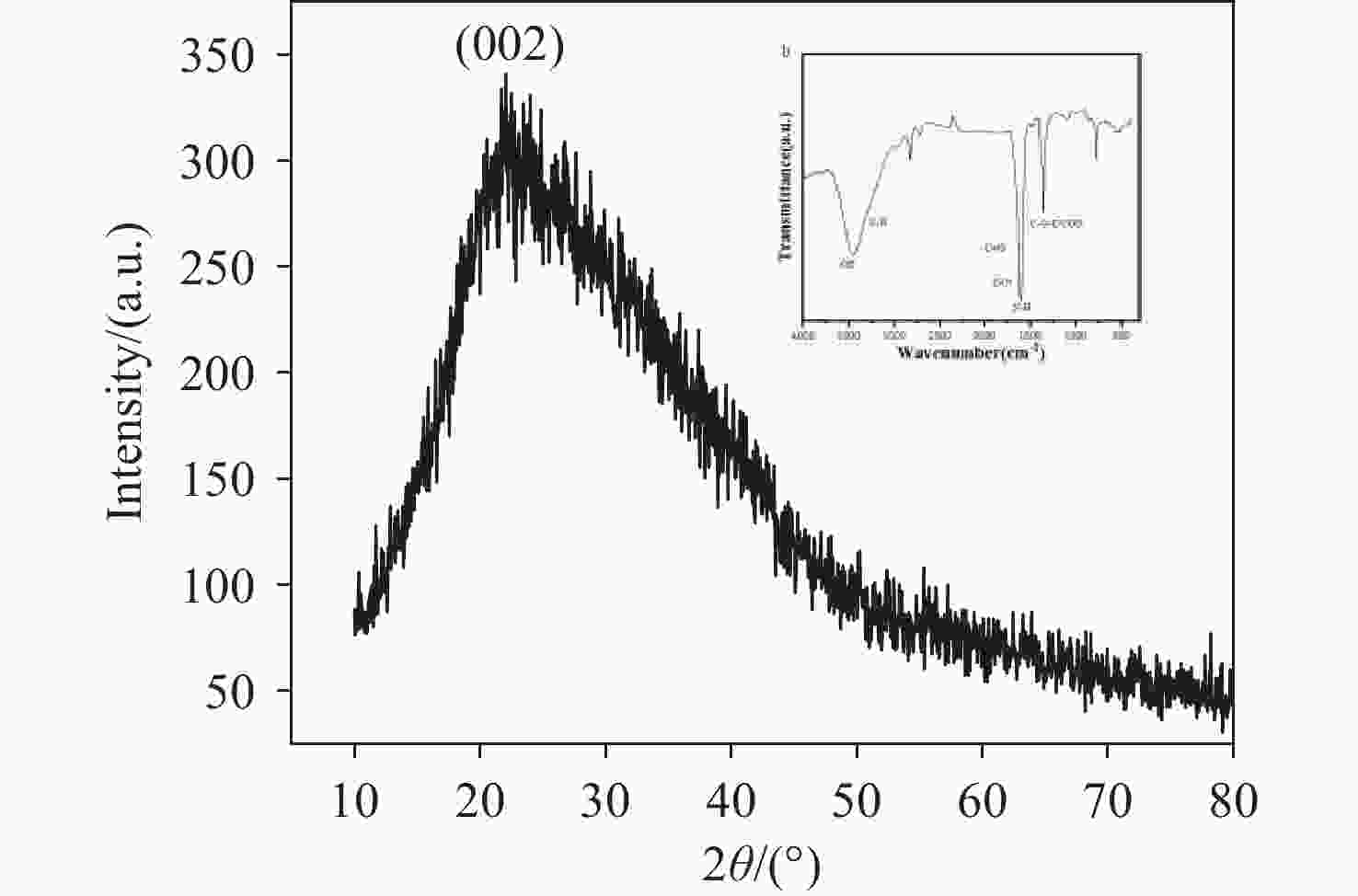
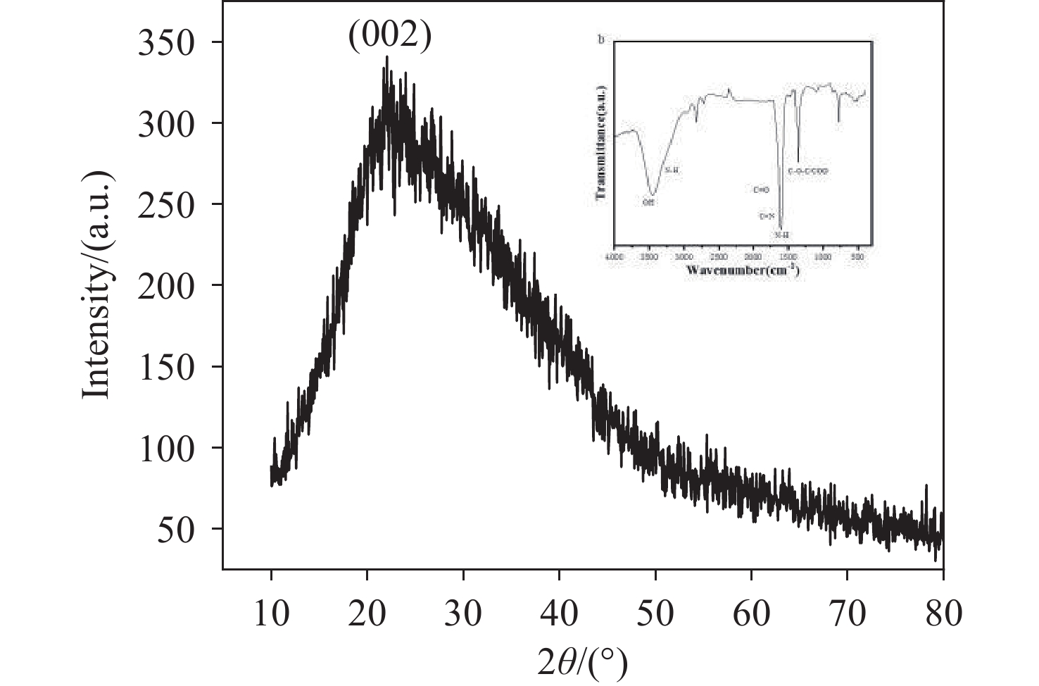
图 2 氮掺杂碳点(N-CDs)的X射线衍射(XRD)图谱, 插图为氮掺杂碳点(N-CDs)的红外光谱
Figure 2. X-ray diffraction (XRD) pattern of the nitrogen-doped carbon dots (N-CDs). The inset shows the corresponding Fourier transform infrared (FTIR) spectrum.
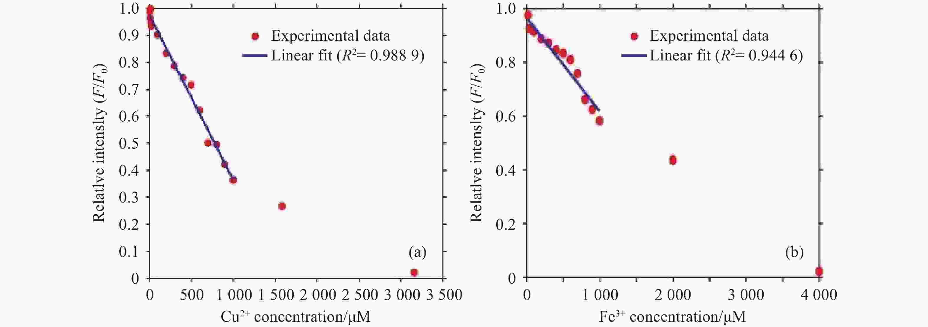
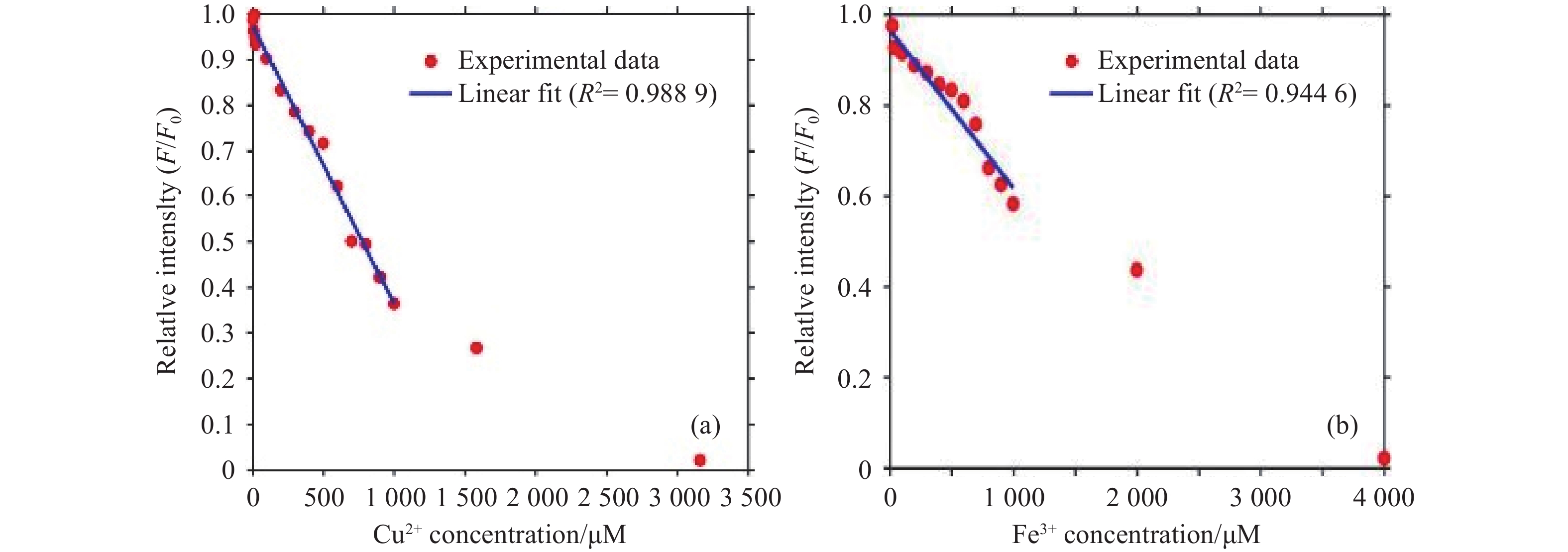
图 3 N-CDs 对单组分金属离子的定量猝灭响应。 (a) 体系相对荧光强度 (F/F0) 随Cu2+浓度的线性响应及检出限拟合曲线;(b) 体系相对荧光强度(F/F0)随Fe3+浓度的线性响应及检出限拟合曲线。
Figure 3. Quantitative quenching response of N-CDs to individual metal ions. Linear fitting curves of the relative fluorescence intensity (F/F0) versus the concentration of (a) Cu2+ and (b) Fe3+ for the determination of limits of detection (LOD).
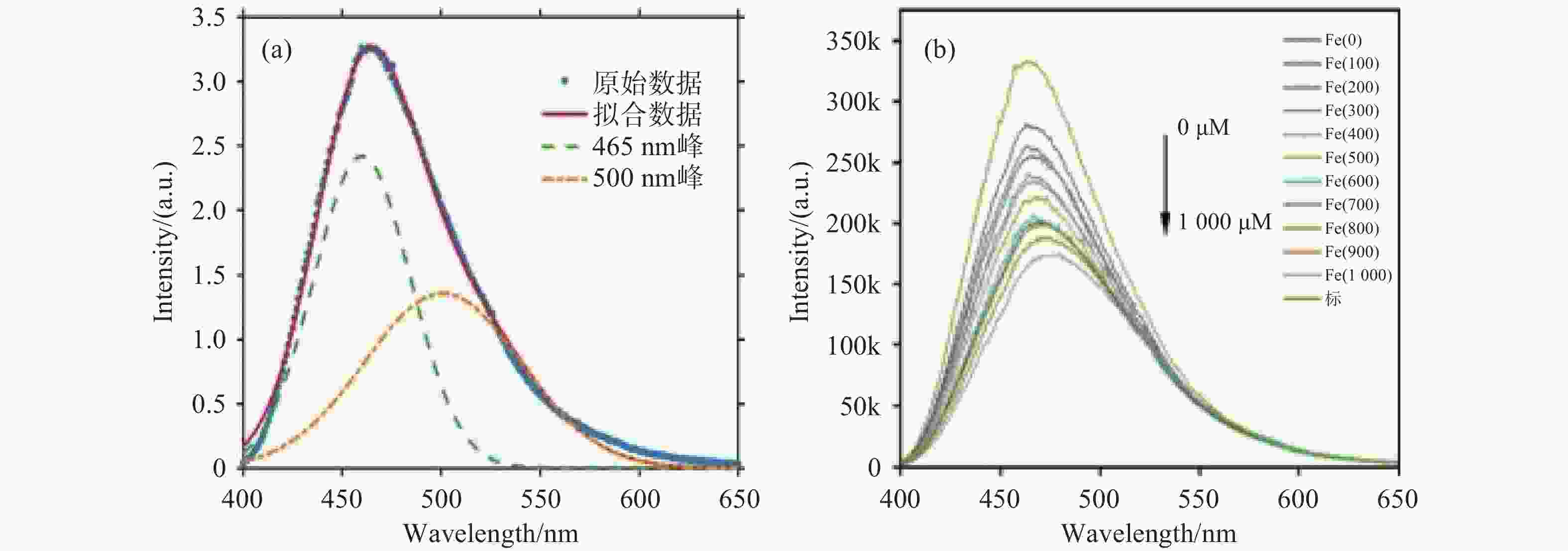
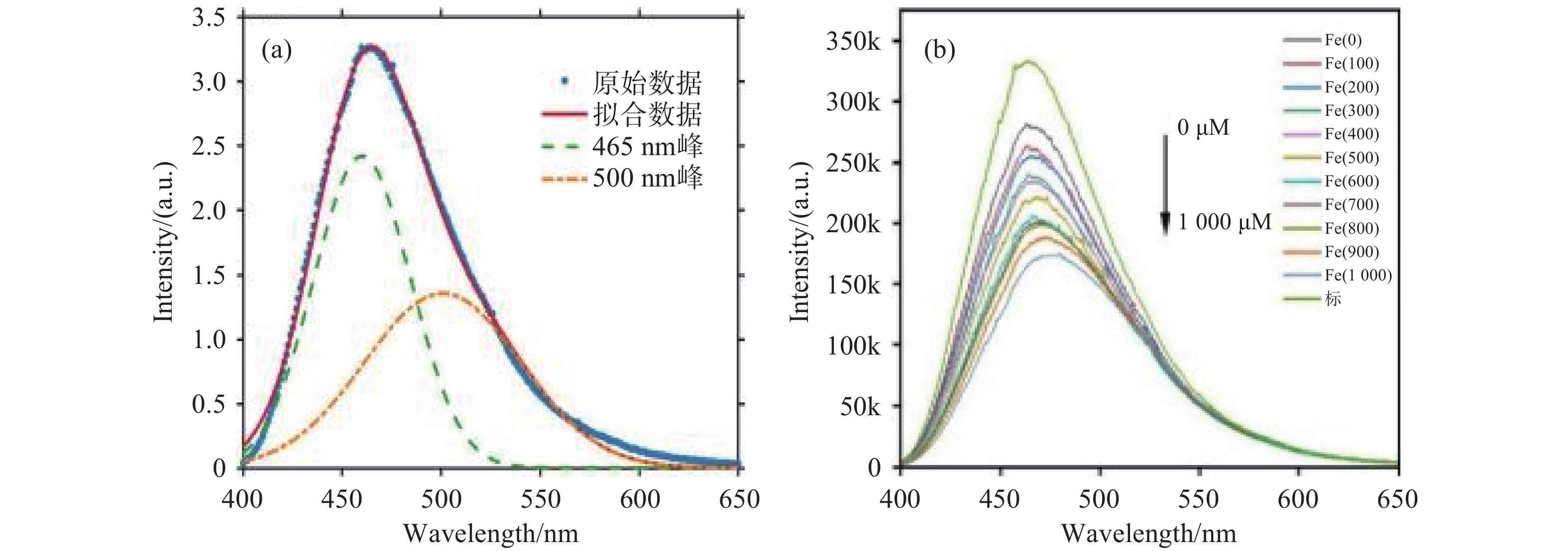
图 4 (a) N-CDs 荧光发射光谱的高斯分峰拟合图(展示了465 nm与500 nm两个发射通道);(b) 固定Cu2+(200 μM) 背景下,Fe3+滴定过程中的荧光光谱演化图
Figure 4. (a) Gaussian peak-fitting of the N-CDs fluorescence emission spectrum, showing the two emission channels at 465 nm and 500 nm; (b) Evolution of fluorescence spectra during Fe3+ titration under a fixed Cu2+ (200 μM) background.
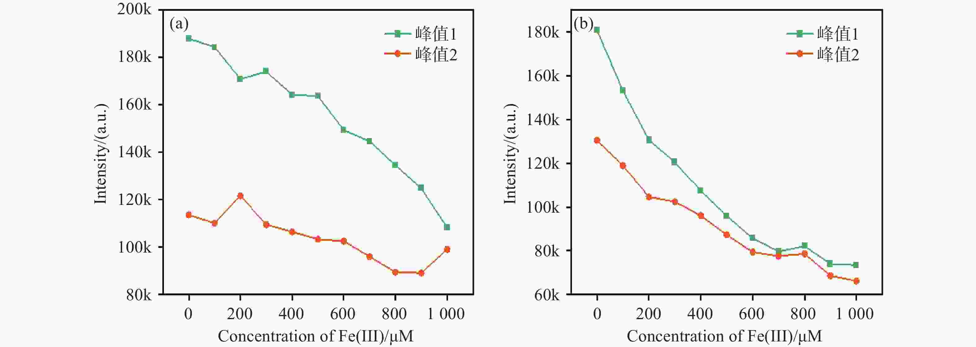
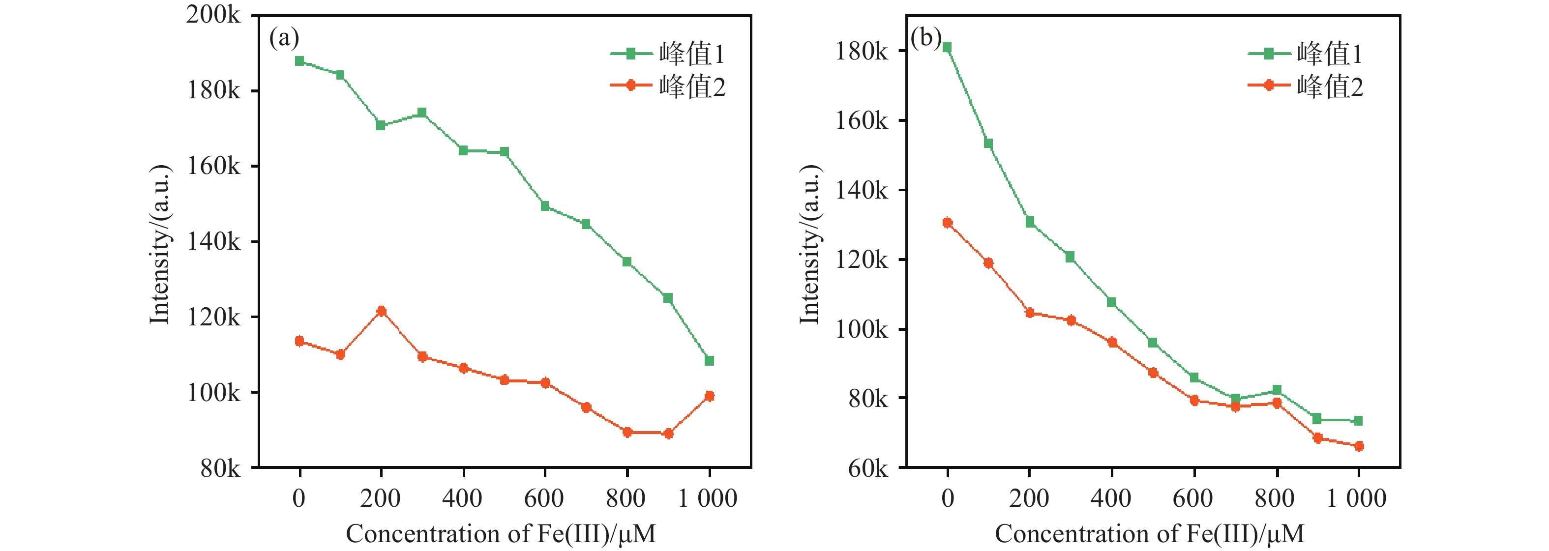
图 5 (a)无干扰背景与(b) 600 μM Cu2+ 强背景下Fe3+ 的荧光猝灭响应曲线
Figure 5. Fluorescence quenching response curves of Fe3+ in (a) an unspiked background and (b) a strong 600 μM Cu2+ background
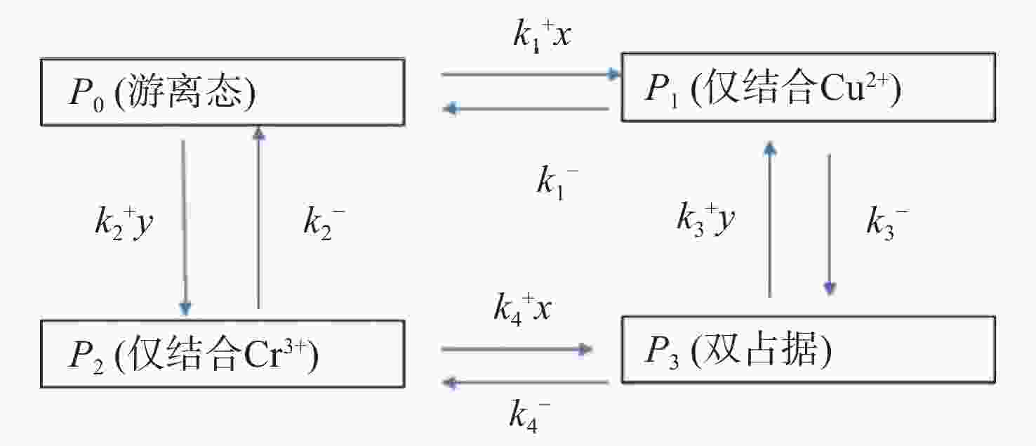
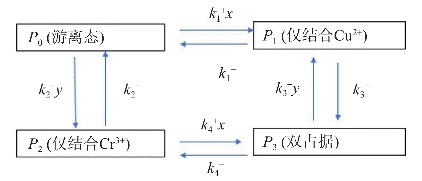
图 6 四种微观状态的空间构型及其稳态分布示意图
Figure 6. Schematic diagram illustrating the spatial configurations and steady-state distributions of the four microscopic states
-
[1] SRIVASTAVA S K, PRATAP R, YADAV M, et al. Biogenic synthesis of highly stable multifluorescent, multifaceted carbon quantum dots as dual probe sensor for detection of heavy toxic metal ions-Fe3+and As3+in water and mouse fibroblast cell line NIH-3T3, and for live in-vivo imaging in mice[J]. Colloid Surf A-Physicochem Eng Asp, 2025, 723: 14. [2] ZHANG L, CAI Z L, LIU Y Q, et al. Fluorescent enhanced endogenous carbon dots derived from green tea residue for multiplex detection of heavy metal ions in food[J]. Front Sustain Food Syst, 2024, 8: 13. [3] YANG X, ZHANG L K, LU B, et al. The green synthesis of low-cost carbon quantum dots as eco-friendly probes in the detection of heavy metal ions[J]. J Mater Sci, 2026, 61(7): 4497-514. doi: 10.1007/s10853-026-12171-7 [4] SHEN Y J, RONG M C, QU X D, et al. Graphene oxide-assisted synthesis of N, S Co-doped carbon quantum dots for fluorescence detection of multiple heavy metal ions[J]. Talanta, 2022, 241: 8. [5] LI S, LI L, TU H, et al. The development of carbon dots: From the perspective of materials chemistry [J]. 2021, 51: 188-207. [6] AI L, YANG Y, WANG B, et al. Insights into photoluminescence mechanisms of carbon dots: advances and perspectives [J]. 2021, 66(8): 839-56. [7] HE C, XU P, ZHANG X, et al. The synthetic strategies, photoluminescence mechanisms and promising applications of carbon dots: Current state and future perspective [J]. 2022, 186: 91-127. [8] SHABBIR H, CSAPó E, WOJNICKI M J I. Carbon quantum dots: the role of surface functional groups and proposed mechanisms for metal ion sensing [J]. 2023, 11(6): 262. [9] YARUR F, MACAIRAN J R, NACCACHE R. Ratiometric detection of heavy metal ions using fluorescent carbon dots[J]. Environmental Science-Nano, 2019, 6(4): 1121-30. doi: 10.1039/C8EN01418C [10] SUN Z, XING H H, QING M, et al. From the perspective of high-throughput recognition: Sulfur quantum dots-based multi-channel sensing platform for metal ions detection [J]. 2023, 452: 139594. [11] MOHANDOSS S, AHMAD N, VELU K S, et al. Synthesis of Photoluminescent Carbon Dots Using Hibiscus Tea Waste and Heteroatom Doping for Multi-Metal Ion Sensing: Applications in Cell imaging and Environmental Samples[J]. Chemosensors, 2023, 11(9): 14. doi: 10.3390/chemosensors11090474 [12] ISSA M A, ABIDIN Z Z, SOBRI S, et al. Fluorescent recognition of Fe3+ in acidic environment by enhanced-quantum yield N-doped carbon dots: optimization of variables using central composite design [J]. 2020, 10(1): 11710. [13] RACHMINA R, HASAN M, SUHARTONO S, et al. Enhanced fluorescence and heavy metal ion sensing using nitrogen-doped carbon quantum dots synthesized from banana peel waste [J]. Results in Engineering, 2026. [14] SAHU G, CHAWRE Y, KUJUR A B, et al. Nitrogen Doped Carbon Quantum Dots as Fluorescence "Turn-Off-On" Sensor for Detection of Fe3+ Ions and Ascorbic Acid in Moringa oleifera and Citrus Lemon[J]. J Fluoresc, 2025, 35(8): 6581-93. [15] MOCCI F, DE VILLIERS ENGELBRECHT L, OLLA C, et al. Carbon nanodots from an in silico perspective [J]. 2022, 122(16): 13709-99. [16] MOHAMMADPOOR M J I C C. Machine learning-driven approaches for synthesizing carbon dots and their applications in photoelectrochemical sensors [J]. 2024, 159: 111859. [17] KURUKAVAK C K, TOK M, TOPRAK A, et al. Effect of Hetero-Atom Doping on the Structure and Optical Properties of Carbon Quantum Dots for the Sensitive Detection of Heavy Metal Ions[J]. Luminescence, 2025, 40(6): 12. doi: 10.1002/bio.70215 [18] ZHANG Y, WANG Y, FENG X, et al. Effect of reaction temperature on structure and fluorescence properties of nitrogen-doped carbon dots [J]. 2016, 387: 1236-46. [19] PUNDI A, CHANG C J. Recent Advances in Synthesis, Modification, Characterization, and Applications of Carbon Dots[J]. Polymers, 2022, 14(11): 40. [20] ÐORĐEVIĆ L, ARCUDI F, CACIOPPO M, et al. A multifunctional chemical toolbox to engineer carbon dots for biomedical and energy applications [J]. 2022, 17(2): 112-30. [21] YAN F, JIANG Y, SUN X, et al. Surface modification and chemical functionalization of carbon dots: a review [J]. 2018, 185(9): 424. [22] NGOC N T B, HA V T H, ANH T N, et al. Rare emission peak shift unlocks detection of multiple heavy metal ions with dual emission carbon dots[J]. Microchem J, 2025, 218: 10. [23] YAO B, HUANG H, LIU Y, et al. Carbon Dots: A Small Conundrum[J]. Trends in Chemistry, 2019, 1(2): 235-46. doi: 10.1016/j.trechm.2019.02.003 [24] SHI L, WANG B, LU S J M. Efficient bottom-up synthesis of graphene quantum dots at an atomically precise level [J]. 2023, 6(3): 728-60. [25] YADAV R, LAHARIYA V, SINGH A K, et al. Fluorometric sensing and nanomolar level detection of heavy metal ions using nitrogen doped carbon dots[J]. Emerg Mater, 2025, 8(1): 363-77. doi: 10.1007/s42247-024-00825-8 [26] HU G K, PEI Z J, SHEN B, et al. Correlation between surface structure of carbon dots and selective detection of heavy metal ions[J]. Appl Phys A-Mater Sci Process, 2024, 130(2): 13. doi: 10.1007/s00339-023-07265-x [27] LI H, YAN X, KONG D, et al. Recent advances in carbon dots for bioimaging applications [J]. 2020, 5(2): 218-34. [28] WANG B, CAI H, WATERHOUSE G I, et al. Carbon dots in bioimaging, biosensing and therapeutics: a comprehensive review [J]. 2022, 2(6): 2200012. [29] KIM N, LEE J, GU M, et al. Modulating charge carriers in carbon dots toward efficient solar‐to‐energy conversion [J]. 2021, 3(4): 590-614. [30] WANG J, FU Y, GU Z, et al. Multifunctional carbon dots for biomedical applications: diagnosis, therapy, and theranostic [J]. 2024, 20(3): 2303773. [31] QIAN H. Cooperativity and specificity in enzyme kinetics: a single-molecule time-based perspective[J]. Biophys J, 2008, 95(1): 10-7. doi: 10.1529/biophysj.108.131771 -


 下载:
下载:

计量
- 文章访问数: 277
- HTML全文浏览量: 72
- PDF下载量: 8
- 被引次数: 0
