


Design of a miniature head-mounted fluorescence microscope based on gradient refractive index lenses
-
摘要:
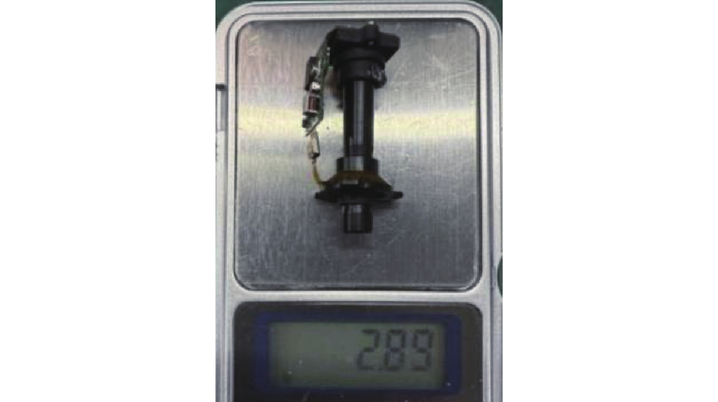
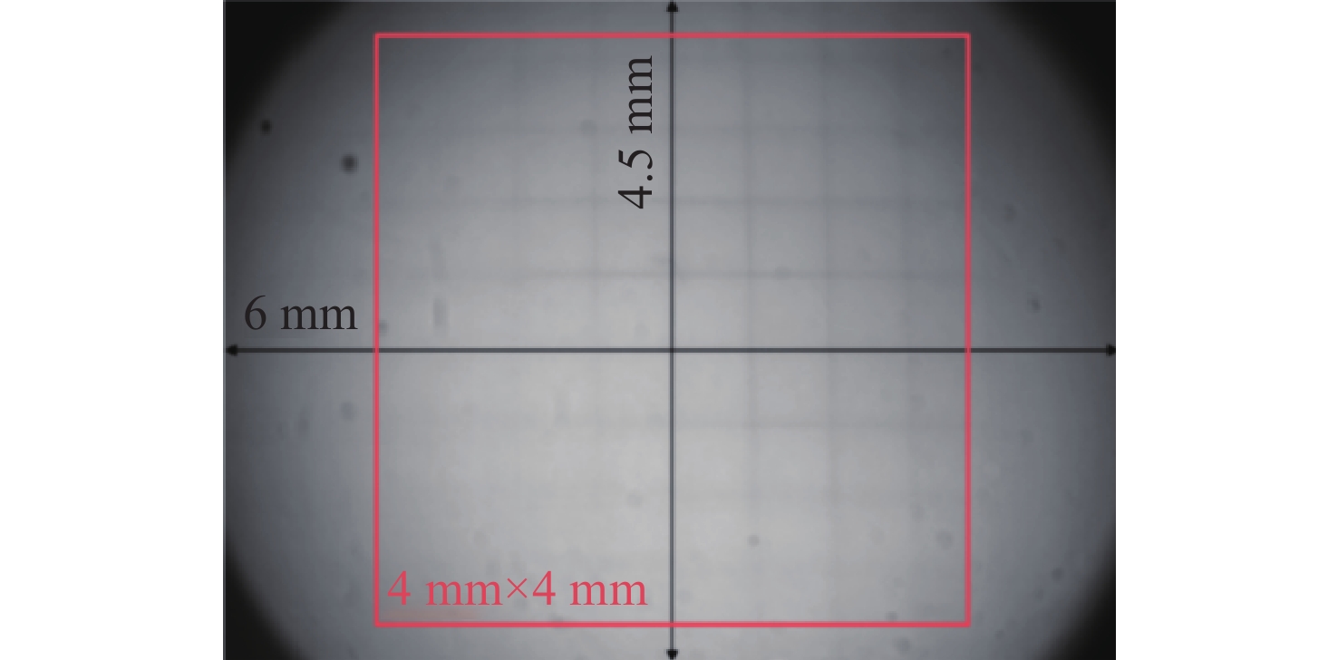
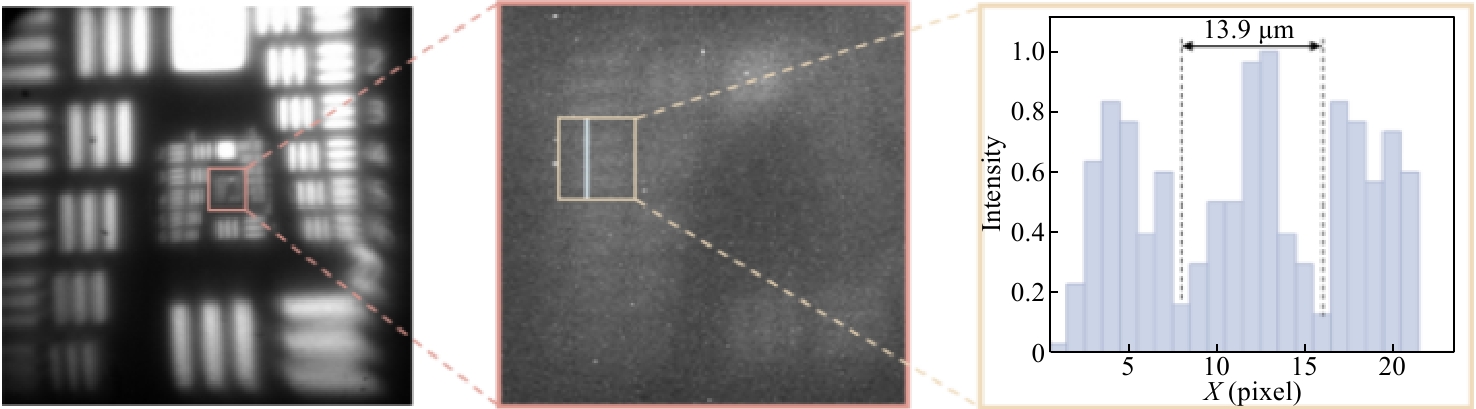
在对自由移动的动物进行脑神经实时观测时,微型头戴式荧光显微镜是当前最为前沿的脑科学观测仪器之一。但是,现今多数微型荧光显微镜为了达到体积和重量的严格限制,使得视场较小,无法同时观测多个脑区的神经活动,而少数视场大的产品重量较大,无法佩戴在小型动物身上。本研究使用轻量化、平面化和成像质量高的梯度折射率透镜,在保证大视场的前提下,减小了显微镜的重量。本文使用梯度折射率透镜进行大视场微型荧光显微镜设计,推导了倾斜光线入射梯度折射率透镜时的离轴像差公式,分析了梯度折射率透镜的折射率排布模型和像差校正情况,并据此设计了一款微型荧光显微镜,其视场为4 mm×4 mm,NA为0.1,样机重量仅为2.89 g,中心视场分辨率为13.9 μm,初步达到了自由移动小鼠的脑神经细胞分辨率。
Abstract:In real-time brain neural observation of freely moving animals, the miniature head-mounted fluorescence microscope is currently one of the most advanced brain science observation instruments. However, most existing miniature fluorescence microscopes, in order to meet strict size and weight constraints, have a limited field of view, making it impossible to simultaneously observe neural activity in multiple brain regions. On the other hand, a few products with a larger field of view are too heavy to be worn on small animals. This study employs lightweight, planar, and high-quality gradient refractive index lenses to reduce the microscope's weight while ensuring a large field of view. Using gradient refractive index lenses for the design of a large-field-of-view miniature fluorescence microscope, this research derives the off-axis aberration formula for oblique light incidence on gradient refractive index lenses, analyzes the refractive index distribution model and aberration correction of these lenses, and designs a miniature fluorescence microscope with a 4 mm×4 mm field of view, a numerical aperture (NA) of 0.1, and a prototype weight of only 2.89 g. The central visual field resolution is 13.9 μm, preliminarily achieving the resolution for neural cells in freely moving mice.
-
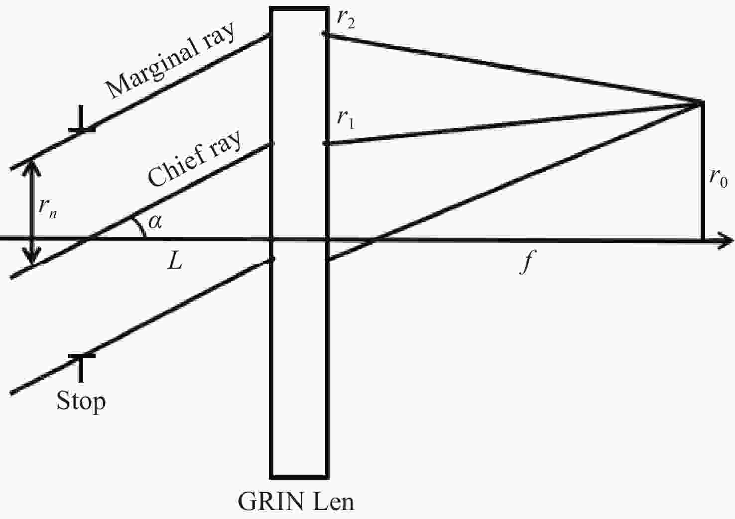
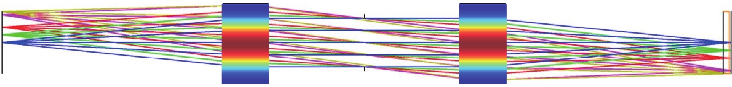
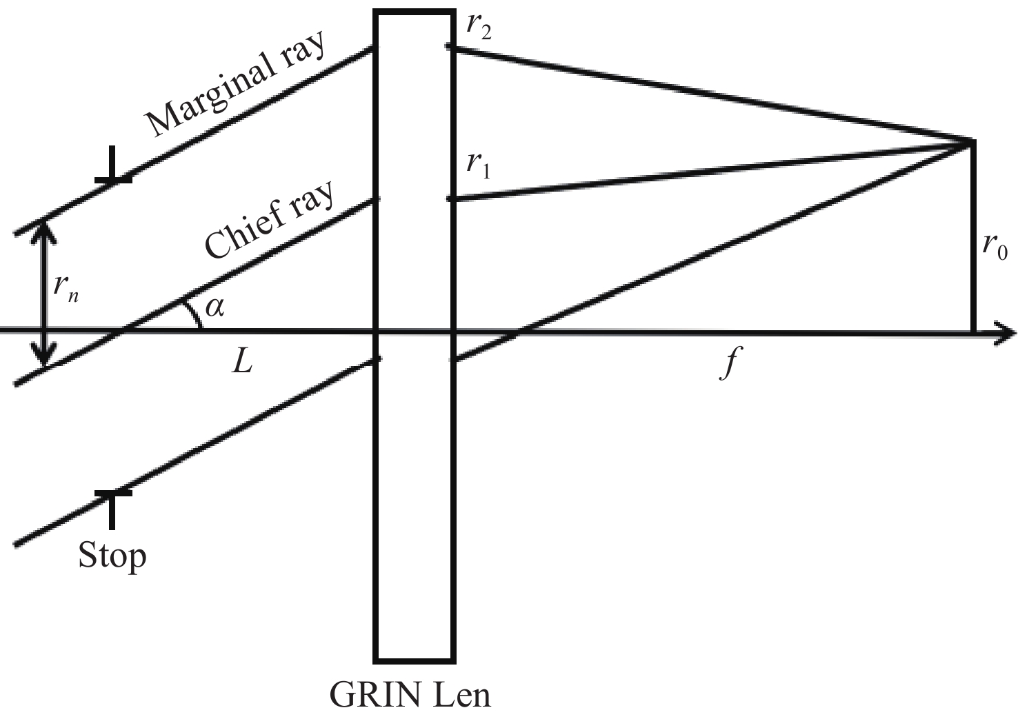
图 1 倾斜光线经过梯度折射率透镜的模拟光路
Figure 1. Simulated optical path of oblique light rays through a gradient refractive index lens
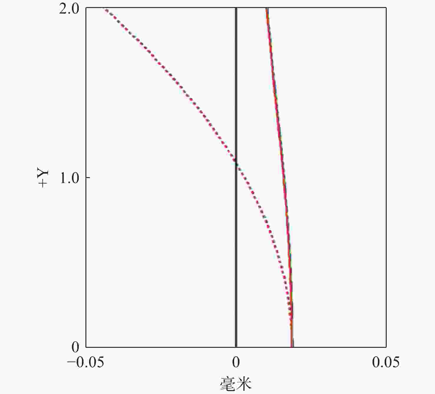
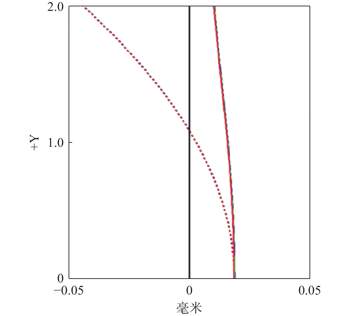
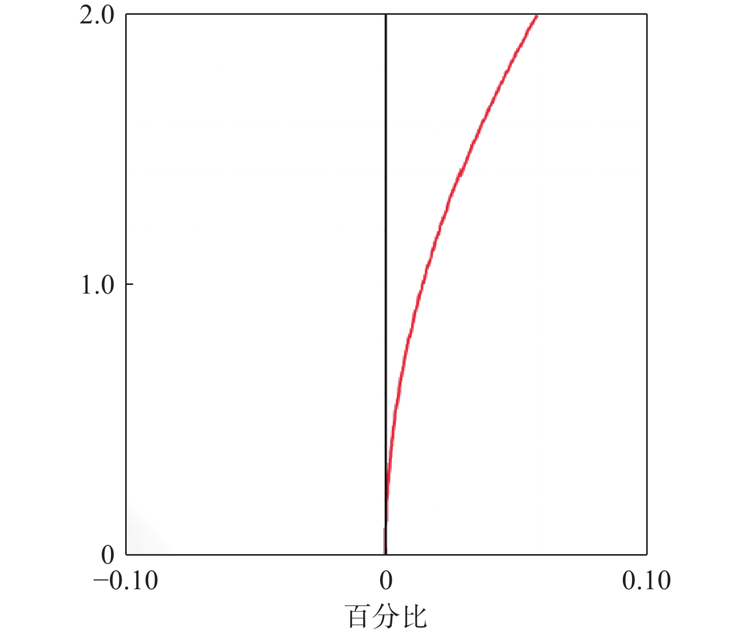
图 6 显微镜的场曲和像散曲线
Figure 6. Field curvature and astigmatism curves of the miniature fluorescence microscope
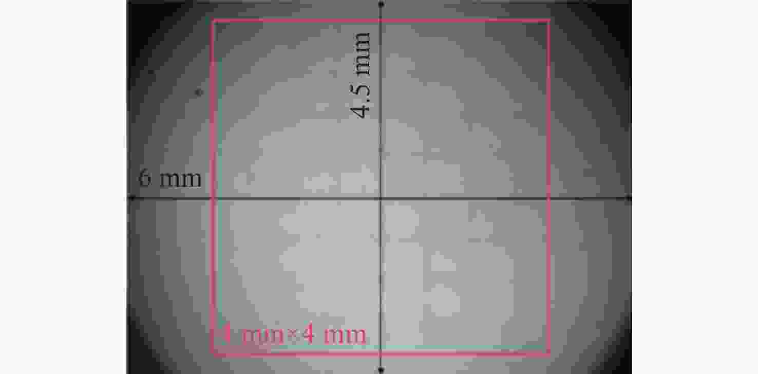
图 11 畸变测试结果
Figure 11. Distortion test results of the miniature fluorescence microscope
-
[1] 刘晓宇, 刘紫千, 斯科, 等. 微型化显微成像系统的关键技术及研究进展(特邀)[J]. 激光与光电子学进展, 2024, 61(2): 0211009. doi: 10.3788/LOP232709LIU X Y, LIU Z Q, SI K, et al. Key technologies and progresses of miniaturized microscopic imaging system (invited)[J]. Laser & Optoelectronics Progress, 2024, 61(2): 0211009. (in Chinese). doi: 10.3788/LOP232709 [2] KAMATH V, BHAT V G, RAJU G, et al. Application of fluorescence lifetime imaging-integrated deep learning analysis for cancer research[J]. Light: Advanced Manufacturing, 2025, 6(3): 49. doi: 10.37188/lam.2025.049 [3] YU H, SENARATHNA J, TYLER B M, et al. Miniaturized optical neuroimaging in unrestrained animals[J]. NeuroImage, 2015, 113: 397-406. doi: 10.1016/j.neuroimage.2015.02.070 [4] CHEN SH Y, WANG Z CH, ZHANG D, et al. Miniature fluorescence microscopy for imaging brain activity in freely-behaving animals[J]. Neuroscience Bulletin, 2020, 36(10): 1182-1190. doi: 10.1007/s12264-020-00561-z [5] HOU Y W, WANG W Y, FU Y ZH, et al. Multi-resolution analysis enables fidelity-ensured deconvolution for fluorescence microscopy[J]. eLight, 2024, 4(1): 14. doi: 10.1186/s43593-024-00073-7 [6] YANNY K, ANTIPA N, LIBERTI W, et al. Miniscope3D: optimized single-shot miniature 3D fluorescence microscopy[J]. Light: Science & Applications, 2020, 9(1): 171. [7] 胡鹏涛, 高若谦, 葛明锋, 等. 流动相单分子免疫检测系统的设计[J]. 中国光学(中英文), 2025, 18(5): 1055-1065. doi: 10.37188/CO.2025-0045HU P T, GAO R Q, GE M F, et al. Design of flow-phase single-molecule immunoassay detection system[J]. Chinese Optics, 2025, 18(5): 1055-1065. (in Chinese). doi: 10.37188/CO.2025-0045 [8] KAMATH V, BHAT V G, RAJU G, et al. Application of fluorescence lifetime imaging-integrated deep learning analysis for cancer research[J]. Light: Advanced Manufacturing, 2025, 6(3): 49. (查阅网上资料, 本条文献与第2条文献重复, 请核对). [9] MOK A T, WANG T Y, ZHAO SH T, et al. A large field-of-view, single-cell-resolution two- and three-photon microscope for deep and wide imaging[J]. eLight, 2024, 4: 20. doi: 10.1186/s43593-024-00076-4 [10] YANNY K, ANTIPA N, LIBERTI W, et al. Miniscope3D: optimized single-shot miniature 3D fluorescence microscopy[J]. Light: Science & Applications, 2020, 9(1): 171. (查阅网上资料, 本条文献与第6条和第24条文献重复, 请核对). [11] 付强, 张智淼, 赵尚男, 等. 微型头戴式单光子荧光显微成像技术研究进展[J]. 中国光学(中英文), 2023, 16(5): 1010-1021.FU Q, ZHANG ZH M, ZHAO SH N, et al. Research progress of miniature head-mounted single photon fluorescence microscopic imaging technique[J]. Chinese Optics, 2023, 16(5): 1010-1021. (in Chinese). [12] RYNES M L, SURINACH D A, LINN S, et al. Miniaturized head-mounted microscope for whole-cortex mesoscale imaging in freely behaving mice[J]. Nature Methods, 2021, 18(4): 417-425. doi: 10.1038/s41592-021-01104-8 [13] PARK D H, JOO B C, CHOI K R, et al. 3D printed miniature attenuated total internal reflection assisted fluorescence microscopy[J]. Scientific Reports, 2025, 15(1): 7683. doi: 10.1038/s41598-025-92204-4 [14] EROFEEV A I, VINOKUROV E K, ANTIFEEV I E, et al. Integration of single-photon miniature fluorescence microscopy and electrophysiological recording methods for in vivo studying hippocampal neuronal activity[J]. J Evol Biochem Phys, 2024, 60(4): 1586-1606. doi: 10.1134/S0022093024040264 [15] GERASIMOV E, PCHITSKAYA E, VLASOVA O, et al. Dynamic changes in the hippocampal neuronal circuits activity following acute stress revealed by miniature fluorescence microscopy imaging[J]. Molecular Brain, 2024, 17(1): 92. doi: 10.1186/s13041-024-01168-5 [16] GERASIMOV E, MITENEV A, PCHITSKAYA E, et al. NeuroActivityToolkit-toolbox for quantitative analysis of miniature fluorescent microscopy data[J]. Journal of Imaging, 2023, 9(11): 243. doi: 10.3390/jimaging9110243 [17] ZHAO P P, GUO CH L, XIE M, et al. MiniXL: An open-source, large field-of-view epifluorescence miniscope enabling single-cell resolution and multi-region imaging in mice[J]. Science Advances, 2025, 11(24): eads4995. doi: 10.1126/sciadv.ads4995 [18] 张智淼, 王承邈, 谢冕, 等. 基于超构透镜的微型头戴式荧光显微镜设计[J]. 中国光学(中英文), 2024, 17(3): 512-520.ZHANG ZH M, WANG CH M, XIE M, et al. Design of miniature head-mounted fluorescence microscope based on metalens[J]. Chinese Optics, 2024, 17(3): 512-520. (in Chinese). [19] SCOTT B B, THIBERGE S Y, GUO C Y, et al. Imaging cortical dynamics in GCaMP transgenic rats with a head-mounted widefield macroscope[J]. Neuron, 2018, 100(5): 1045-1058. e5. [20] RYNES M L, SURINACH D A, LINN S, et al. Miniaturized head-mounted microscope for whole-cortex mesoscale imaging in freely behaving mice[J]. Nature Methods, 2021, 18(4): 417-425. (查阅网上资料, 本条文献与第12条文献重复, 请核对). [21] SUPEKAR O D, SIAS A, HANSEN S R, et al. Miniature structured illumination microscope for in vivo 3D imaging of brain structures with optical sectioning[J]. Biomedical Optics Express, 2022, 13(4): 2530-2541. doi: 10.1364/BOE.449533 [22] GUO CH L, BLAIR G J, SEHGAL M, et al. Miniscope-LFOV: a large-field-of-view, single-cell-resolution, miniature microscope for wired and wire-free imaging of neural dynamics in freely behaving animals[J]. Science Advances, 2023, 9(16): eadg3918. doi: 10.1126/sciadv.adg3918 [23] BAGRAMYAN A. Lightweight 1-photon miniscope for imaging in freely behaving animals at subcellular resolution[J]. IEEE Photonics Technology Letters, 2020, 32(15): 909-912. doi: 10.1109/LPT.2020.3004283 [24] YANNY K, ANTIPA N, LIBERTI W, et al. Miniscope3D: optimized single-shot miniature 3D fluorescence microscopy[J]. Light: Science & Applications, 2020, 9(1): 171. (查阅网上资料, 本条文献与第6条和第10条文献重复, 请核对). [25] WANG Y ZH, MA ZH T, LI W ZH, et al. Cable-free brain imaging for multiple free-moving animals with miniature wireless microscopes[J]. Journal of Biomedical Optics, 2023, 28(2): 026503. doi: 10.1117/1.jbo.28.2.026503 [26] XUE F, LI F, ZHANG K M, et al. Multi-region calcium imaging in freely behaving mice with ultra-compact head-mounted fluorescence microscopes[J]. National Science Review, 2024, 11(1): nwad294. doi: 10.1101/2023.10.30.564709 [27] YANG T Y. Freeform gradient-index optics with applications in rotationally variant systems[D]. Rochester: University of Rochester, 2022. [28] DESAI A X. Chromatic properties of freeform and multi-material gradient-index optics[D]. Rochester: University of Rochester, 2019. (查阅网上资料, 未找到本条文献信息, 请确认). [29] YOUNG M. Zone plates and their aberrations[J]. Journal of the Optical Society of America, 1972, 62(8): 972-976. doi: 10.1364/JOSA.62.000972 [30] GROSS H. Handbook of Optical Systems (Volume 3: Aberration Theory and Correction of Optical Systems)[M]. Weinheim: John Wiley & Sons Inc., 2007. -

 下载:
下载:

计量
- 文章访问数: 100
- HTML全文浏览量: 51
- PDF下载量: 3
- 被引次数: 0
