| Citation: | WANG De-jiang, DI Xiang-jun, WANG Bao-ming, WANG Fan, GUO Zhi-yong, JIN Da-yong. Advances in single particle tracking in living cells[J]. Chinese Optics, 2018, 11(3): 281-295. doi: 10.3788/CO.20181103.0281

|

| [1] |
LIPPINCOTT-SCHWARTZ J, SNAPP E, KENWORTHY A. Studying protein dynamics in living cells[J]. Nat. Rev. Mol. Cell Bio., 2001, 2(6):444-456. doi: 10.1038/35073068
|
| [2] |
REITS E A J, NEEFJES J J. From fixed to FRAP:measuring protein mobility and activity in living cells[J]. Nat. Cell Biol., 20013(6):E145-E147. doi: 10.1038/35078615
|
| [3] |
DOMINGUEZ-MEDINA S, CHEN S, BLANKENBURG J, et al.. Measuring the hydrodynamic size of nanoparticles using fluctuation correlation spectroscopy[J]. Annual Review of Physical Chemistry, 2016, 67:489-514. doi: 10.1146/annurev-physchem-040214-121510
|
| [4] |
YU TA, MARTINEZ MM, PAPPAS D. Fluorescence correlation spectroscopy:a review of biochemical and microfluidic applications[J]. Applied Spectroscopy, 2011, 65(4):115a-124a. doi: 10.1366/10-06224
|
| [5] |
SPRAGUE B L, PEGO R L, STAVREVA DA, et al.. Analysis of binding reactions by fluorescence recovery after photobleaching[J]. Biophysical Journal, 2004, 86(6):3473-3495. doi: 10.1529/biophysj.103.026765
|
| [6] |
ELSON E L. Fluorescence correlation spectroscopy:past, present, future[J]. Biophysical Journal, 2011, 101(12):2855-2870. doi: 10.1016/j.bpj.2011.11.012
|
| [7] |
HAUSTEIN E, SCHWILLE P. Fluorescence correlation spectroscopy:novel variations of an established technique[J]. Annual Review of Biophysics and Biomolecular Structure, 2007, 36:151-169. doi: 10.1146/annurev.biophys.36.040306.132612
|
| [8] |
GEERTS H, DE BRABANDER M, NUYDENS R, et al.. Nanovid tracking:a new automatic method for the study of mobility in living cells based on colloidal gold and video microscopy[J]. Biophysical Journal, 1987, 52(5):775-782. doi: 10.1016/S0006-3495(87)83271-X
|
| [9] |
DE BRABANDER M, NUYDENS R, GEERTS H, et al.. Dynamic behavior of the transferrin receptor followed in living epidermoid carcinoma(A431) cells with nanovid microscopy[J]. Cell Motility and the Cytoskeleton, 1988, 9(1):30-47. doi: 10.1002/(ISSN)1097-0169
|
| [10] |
DE BRABANDER M, NUYDENS R, ISHIHARA A, et al.. Lateral diffusion and retrograde movements of individual cell surface components on single motile cells observed with Nanovid microscopy[J]. The Journal of Cell Biology, 1991, 112(1):111-124. doi: 10.1083/jcb.112.1.111
|
| [11] |
BETZIG E, CHICHESTER R J. Single molecules observed by near-field scanning optical microscopy[J]. Science, 1993, 262(5138):1422-1425. doi: 10.1126/science.262.5138.1422
|
| [12] |
SAKO Y, MINOGHCHI S, YANAGIDA T. Single-molecule imaging of EGFR signalling on the surface of living cells[J]. Nature Cell Biology, 2000, 2(3):168-172. doi: 10.1038/35004044
|
| [13] |
ⅡNO R, KOYAMA I, KUSUMI A. Single molecule imaging of green fluorescent proteins in living cells:E-cadherin forms oligomers on the free cell surface[J]. Biophysical Journal, 2001, 80(6):2667-2677. http://www.cell.com/biophysj/pdf/S0006-3495(01)76236-4.pdf
|
| [14] |
SCHMIDT T, SCHUTZ GJ, BAUMGARTNER W, et al.. Imaging of single molecule diffusion[J]. Proceedings of the National Academy of Sciences of the United States of America, 1996, 93(7):2926-2929. doi: 10.1073/pnas.93.7.2926
|
| [15] |
SCHUTZ G J, KADA G, PASTUSHENKO V P, et al.. Properties of lipid microdomains in a muscle cell membrane visualized by single molecule microscopy[J]. The EMBO Journal, 2000, 19(5):892-901. doi: 10.1093/emboj/19.5.892
|
| [16] |
DAHAN M, LEVI S, LUCCARDINI C, et al.. Diffusion dynamics of glycine receptors revealed by single-quantum dot tracking[J]. Science, 2003, 302(5644):442-445. doi: 10.1126/science.1088525
|
| [17] |
KUSUMI A, SUZUKI K G, KASAI R S, et al.. Hierarchical mesoscale domain organization of the plasma membrane[J]. Trends in Biochemical Sciences, 2011, 36(11):604-615. doi: 10.1016/j.tibs.2011.08.001
|
| [18] |
WARSHAW D M, KENNEDY G G, WORK S S, et al.. Differential labeling of myosin V heads with quantum dots allows direct visualization of hand-over-hand processivity[J]. Biophysical Journal, 2005, 88(5):L30-32. doi: 10.1529/biophysj.105.061903
|
| [19] |
NAN X, SIMS P A, CHEN P, et al.. Observation of individual microtubule motor steps in living cells with endocytosed quantum dots[J]. The Journal of Physical Chemistry B, 2005, 109(51):24220-24224. doi: 10.1021/jp056360w
|
| [20] |
COURTY S, LUCCARDINI C, BELLAICHE Y, et al.. Tracking individual kinesin motors in living cells using single quantum-dot imaging[J]. Nano Letters, 2006, 6(7):1491-1495. doi: 10.1021/nl060921t
|
| [21] |
BIERMANN B, SOKOLL S, KLUEVA J, et al.. Imaging of molecular surface dynamics in brain slices using single-particle tracking[J]. Nature Communications, 2014, 5. http://cn.bing.com/academic/profile?id=c801e105fb5de70e3ee952400841fee7&encoded=0&v=paper_preview&mkt=zh-cn
|
| [22] |
NAM S H, BAE Y M, PARK Y I, et al.. Long-term real-time tracking of lanthanide ion doped upconverting nanoparticles in living cells[J]. Angewandte Chemie, 2011, 50(27):6093-6097. doi: 10.1002/anie.v50.27
|
| [23] |
KURAL C, KIM H, SYED S, et al.. Kinesin and dynein move a peroxisome in vivo:a tug-of-war or coordinated movement?[J]. Science, 2005, 308(5727):1469-1472. doi: 10.1126/science.1108408
|
| [24] |
YILDIZ A, FORKEY J N, MCKINNEY S A, et al.. Myosin V walks hand-over-hand:single fluorophore imaging with 1.5-nm localization[J]. Science, 2003, 300(5628):2061-2065. doi: 10.1126/science.1084398
|
| [25] |
CHEN K, GU Y, SUN W, et al.. Characteristic rotational behaviors of rod-shaped cargo revealed by automated five-dimensional single particle tracking[J]. Nature Communications, 2017, 8(1):887. doi: 10.1038/s41467-017-01001-9
|
| [26] |
CHEEZUM M K, WALKER W F, GUILFORD W H. Quantitative comparison of algorithms for tracking single fluorescent particles[J]. Biophysical Journal, 2001, 81(4):2378-2388. doi: 10.1016/S0006-3495(01)75884-5
|
| [27] |
SHEN H, TAUZIN L J, BAIYASI R, et al.. Single particle tracking:from theory to biophysical applications[J]. Chemical Reviews, 2017, 117(11):7331-7376. doi: 10.1021/acs.chemrev.6b00815
|
| [28] |
MANZO C, GARCIA-PARAJO M F. A review of progress in single particle tracking:from methods to biophysical insights[J]. Reports on Progress in Physics. Physical Society, 2015, 78(12):124601. doi: 10.1088/0034-4885/78/12/124601
|
| [29] |
SERGE A, BERTAUX N, RIGNEAULT H, et al.. Dynamic multiple-target tracing to probe spatiotemporal cartography of cell membranes[J]. Nature Methods, 2008, 5(8):687-694. doi: 10.1038/nmeth.1233
|
| [30] |
SAXTON M J, JACOBSON K. Single-particle tracking:applications to membrane dynamics[J]. Annual Review of Biophysics and Biomolecular Structure, 1997, 26:373-399. doi: 10.1146/annurev.biophys.26.1.373
|
| [31] |
DEAN K M, PALMER A E. Advances in fluorescence labeling strategies for dynamic cellular imaging[J]. Nature Chemical Biology, 2014, 10(7):512-523. doi: 10.1038/nchembio.1556
|
| [32] |
FERNANDEZ-SUAREZ M, TING A Y. Fluorescent probes for super-resolution imaging in living cells[J]. Nature Reviews. Molecular Cell Biology, 2008, 9(12):929-943. doi: 10.1038/nrm2531
|
| [33] |
SHEETZ M P, TURNEY S, QIAN H, et al.. Nanometre-level analysis demonstrates that lipid flow does not drive membrane glycoprotein movements[J]. Nature, 1989, 340(6231):284-288. doi: 10.1038/340284a0
|
| [34] |
GARCIA-PARAJO M F, SEGERS-NOLTEN G M, VEERMAN J A, et al.. Real-time light-driven dynamics of the fluorescence emission in single green fluorescent protein molecules[J]. Proceedings of the National Academy of Sciences of the United States of America, 2000, 97(13):7237-7242. doi: 10.1073/pnas.97.13.7237
|
| [35] |
BRUCHEZ M J R, MORONNE M, GIN P, et al. Semiconductor nanocrystals as fluorescent biological labels[J]. Science, 1998, 281(5385):2013-2016. doi: 10.1126/science.281.5385.2013
|
| [36] |
YAO J, LARSON D R, VISHWASRAO H D, et al.. Blinking and nonradiant dark fraction of water-soluble quantum dots in aqueous solution[J]. Proceedings of the National Academy of Sciences of the United States of America, 2005, 102(40):14284-14289. doi: 10.1073/pnas.0506523102
|
| [37] |
DERFUS A M, CHAN W C W, BHATIA S N. Probing the cytotoxicity of semiconductor quantum dots[J]. Nano Letters, 2004, 4(1):11-18. doi: 10.1021/nl0347334
|
| [38] |
MIAO P, HAN K, TANG Y G, et al.. Recent advances in carbon nanodots:synthesis, properties and biomedical applications[J]. Nanoscale, 2015, 7(5):1586-1595. doi: 10.1039/C4NR05712K
|
| [39] |
MIAO P, TANG Y G, HANA K, et al.. Facile synthesis of carbon nanodots from ethanol and their application in ferric(Ⅲ) ion assay[J]. J. Mater. Chem. A, 2015, 3(29):15068-15073. doi: 10.1039/C5TA03278D
|
| [40] |
CHANG Y R, LEE H Y, CHEN K, et al.. Mass production and dynamic imaging of fluorescent nanodiamonds[J]. Nature Nanotechnology, 2008, 3(5):284-288. doi: 10.1038/nnano.2008.99
|
| [41] |
FU C C, LEE H Y, CHEN K, et al.. Characterization and application of single fluorescent nanodiamonds as cellular biomarkers[J]. Proceedings of the National Academy of Sciences of the United States of America, 2007, 104(3):727-732. doi: 10.1073/pnas.0605409104
|
| [42] |
JIN H, HELLER D A, STRANO M S. Single-particle tracking of endocytosis and exocytosis of single-walled carbon nanotubes in NIH-3T3 cells[J]. Nano Letters, 2008, 8(6):1577-1585. doi: 10.1021/nl072969s
|
| [43] |
JIN H, HELLER D A, SHARMA R, et al. Size-dependent cellular uptake and expulsion of single-walled carbon nanotubes:single particle tracking and a generic uptake model for nanoparticles[J]. ACS Nano, 2009, 3(1):149-158. doi: 10.1021/nn800532m
|
| [44] |
WU S, HAN G, MILLIRON D J, et al.. Non-blinking and photostable upconverted luminescence from single lanthanide-doped nanocrystals[J]. Proceedings of the National Academy of Sciences of the United States of America, 2009, 106(27):10917-10921. doi: 10.1073/pnas.0904792106
|
| [45] |
BAE Y M, PARK Y I, NAM S H, et al.. Endocytosis, intracellular transport, and exocytosis of lanthanide-doped upconverting nanoparticles in single living cells[J]. Biomaterials, 2012, 33(35):9080-9086. doi: 10.1016/j.biomaterials.2012.08.039
|
| [46] |
WANG F, HAN Y, LIM C S, et al.. Simultaneous phase and size control of upconversion nanocrystals through lanthanide doping[J]. Nature, 2010, 463(7284):1061-1065. doi: 10.1038/nature08777
|
| [47] |
LIU X J, TU Y, GAI H W. Imaging of single molecules by wide-field optical microscopy[J]. Prog. Chem., 2013, 25(2-3):370-379. doi: 10.7536/PC120842
|
| [48] |
MICHALET X, COLYER R A, SCALIA G, et al.. Development of new photon-counting detectors for single-molecule fluorescence microscopy[J]. Philos. T. R. Soc. B, 2013, 368(1611) http://cn.bing.com/academic/profile?id=aae4dbdd448b3ebf29f19710702ad390&encoded=0&v=paper_preview&mkt=zh-cn
|
| [49] |
WIESER S, SCHUTZ G J. Tracking single molecules in the live cell plasma membrane-Do's and Don't's[J]. Methods, 2008, 46(2):131-140. doi: 10.1016/j.ymeth.2008.06.010
|
| [50] |
MICHALET X, SIEGMUND O H W, VALLERGA J V, et al.. Detectors for single-molecule fluorescence imaging and spectroscopy[J]. J. Mod. Optic, 2007, 54(2-3):239-281. doi: 10.1080/09500340600769067
|
| [51] |
AXELROD D. Total internal reflection fluorescence microscopy in cell biology[J]. Traffic, 2001, 2(11):764-774. doi: 10.1034/j.1600-0854.2001.21104.x
|
| [52] |
TOKUNAGA M, IMAMOTO N, SAKATA-SOGAWA K. Highly inclined thin illumination enables clear single-molecule imaging in cells[J]. Nature Methods, 2008, 5(2):159-161. doi: 10.1038/nmeth1171
|
| [53] |
STENDER A S, MARCHUK K, LIU C, et al.. Single cell optical imaging and spectroscopy[J]. Chemical Reviews, 2013, 113(4):2469-2527. doi: 10.1021/cr300336e
|
| [54] |
ARHEL N, GENOVESIO A, KIM K A, et al.. Quantitative four-dimensional tracking of cytoplasmic and nuclear HIV-1 complexes[J]. Nature Methods, 2006, 3(10):817-824. doi: 10.1038/nmeth928
|
| [55] |
LANGE S, KATAYAMA Y, SCHMID M, et al.. Simultaneous transport of different localized mRNA species revealed by live-cell imaging[J]. Traffic, 2008, 9(8):1256-1267. doi: 10.1111/tra.2008.9.issue-8
|
| [56] |
MANLEY S, GILLETTE J M, PATTERSON G H, et al.. High-density mapping of single-molecule trajectories with photoactivated localization microscopy[J]. Nature Methods, 2008, 5(2):155-157. doi: 10.1038/nmeth.1176
|
| [57] |
PASZEK M J, DUFORT C C, ROSSIER O, et al.. The cancer glycocalyx mechanically primes integrin-mediated growth and survival[J]. Nature, 2014, 511(7509):319-325. doi: 10.1038/nature13535
|
| [58] |
ROSSIER O, OCTEAU V, SIBARITA J B, et al.. Integrins beta1 and beta3 exhibit distinct dynamic nanoscale organizations inside focal adhesions[J]. Nature Cell Biology, 2012, 14(10):1057-1067. doi: 10.1038/ncb2588
|
| [59] |
GIANNONE G, HOSY E, LEVET F, et al.. Dynamic superresolution imaging of endogenous proteins on living cells at ultra-high density[J]. Biophysical Journal, 2010, 99(4):1303-1310. doi: 10.1016/j.bpj.2010.06.005
|
| [60] |
GARCIA-PARAJO M F, CAMBI A, TORRENO-PINA J A, et al.. Nanoclustering as a dominant feature of plasma membrane organization[J]. Journal of Cell Science, 2014, 127(Pt 23):4995-5005. http://cn.bing.com/academic/profile?id=9eda786cb0e24492fe6eb11becba9b10&encoded=0&v=paper_preview&mkt=zh-cn
|
| [61] |
LINGWOOD D, SIMONS K. Lipid rafts as a membrane-organizing principle[J]. Science, 2010, 327(5961):46-50. doi: 10.1126/science.1174621
|
| [62] |
KUSUMI A, TSUNOYAMA T A, HIROSAWA K M, et al.. Tracking single molecules at work in living cells[J]. Nature Chemical Biology, 2014, 10(7):524-532. doi: 10.1038/nchembio.1558
|
| [63] |
KUSUMI A, NAKADA C, RITCHIE K, et al.. Paradigm shift of the plasma membrane concept from the two-dimensional continuum fluid to the partitioned fluid:high-speed single-molecule tracking of membrane molecules[J]. Annual Review of Biophysics and Biomolecular Structure, 2005, 34:351-378. doi: 10.1146/annurev.biophys.34.040204.144637
|
| [64] |
SAKO Y, KUSUMI A. Barriers for lateral diffusion of transferrin receptor in the plasma membrane as characterized by receptor dragging by laser tweezers:fence versus tether[J]. The Journal of Cell Biology, 1995, 129(6):1559-1574. doi: 10.1083/jcb.129.6.1559
|
| [65] |
FUJIWARA T, RITCHIE K, MURAKOSHI H, et al.. Phospholipids undergo hop diffusion in compartmentalized cell membrane[J]. The Journal of Cell Biology, 2002, 157(6):1071-1081. doi: 10.1083/jcb.200202050
|
| [66] |
SUZUKI K G, KASAI R S, HIROSAWA K M, et al.. Transient GPI-anchored protein homodimers are units for raft organization and function[J]. Nature Chemical Biology, 2012, 8(9):774-783. doi: 10.1038/nchembio.1028
|
| [67] |
ANDREWS N L, LIDKE K A, PFEIFFER J R, et al.. Actin restricts FcepsilonRI diffusion and facilitates antigen-induced receptor immobilization[J]. Nature Cell Biology, 2008, 10(8):955-963. doi: 10.1038/ncb1755
|
| [68] |
OWEN D M, WILLIAMSON D, RENTERO C, et al.. Quantitative microscopy:protein dynamics and membrane organisation[J]. Traffic, 2009, 10(8):962-971. doi: 10.1111/tra.2009.10.issue-8
|
| [69] |
TREANOR B, DEPOIL D, GONZALEZ-GRANJA A, et al.. The membrane skeleton controls diffusion dynamics and signaling through the B cell receptor[J]. Immunity, 2010, 32(2):187-199. doi: 10.1016/j.immuni.2009.12.005
|
| [70] |
MURASE K, FUJIWARA T, UMEMURA Y, et al.. Ultrafine membrane compartments for molecular diffusion as revealed by single molecule techniques[J]. Biophysical Journal, 2004, 86(6):4075-4093. doi: 10.1529/biophysj.103.035717
|
| [71] |
WIESER S, MOERTELMAIER M, FUERTBAUER E, et al.. (Un)confined diffusion of CD59 in the plasma membrane determined by high-resolution single molecule microscopy[J]. Biophysical Journal, 2007, 92(10):3719-3728. doi: 10.1529/biophysj.106.095398
|
| [72] |
WEGNER K D, HILDEBRANDT N. Quantum dots:bright and versatile in vitro and in vivo fluorescence imaging biosensors[J]. Chem. Soc. Rev., 2015, 44(14):4792-4834. doi: 10.1039/C4CS00532E
|
| [73] |
LEDUC C, SI S, GAUTIER J, et al.. A highly specific gold nanoprobe for live-cell single-molecule imaging[J]. Nano Letters, 2013, 13(4):1489-1494. doi: 10.1021/nl304561g
|
| [74] |
TAURAN Y, BRIOUDE A, COLEMAN A W, et al.. Molecular recognition by gold, silver and copper nanoparticles[J]. World Journal of Biological Chemistry, 2013, 4(3):35-63. doi: 10.4331/wjbc.v4.i3.35
|
| [75] |
LIDKE DS, LIDKE KA, RIEGER B, et al.. Reaching out for signals:filopodia sense EGF and respond by directed retrograde transport of activated receptors[J]. The Journal of Cell Biology, 2005, 170(4):619-626. doi: 10.1083/jcb.200503140
|
| [76] |
RAJAN S S, LIU H Y, VU T Q. Ligand-bound quantum dot probes for studying the molecular scale dynamics of receptor endocytic trafficking in live cells[J]. ACS Nano, 2008, 2(6):1153-1166. doi: 10.1021/nn700399e
|
| [77] |
BHATIA D, ARUMUGAM S, NASILOWSKI M, et al.. Quantum dot-loaded monofunctionalized DNA icosahedra for single-particle tracking of endocytic pathways[J]. Nature Nanotechnology, 2016, 11(12):1112-1119. doi: 10.1038/nnano.2016.150
|
| [78] |
PIEROBON P, ACHOURI S, COURTY S, et al.. Velocity, processivity, and individual steps of single myosin V molecules in live cells[J]. Biophysical Journal, 2009, 96(10):4268-4275. doi: 10.1016/j.bpj.2009.02.045
|
| [79] |
FAKHRI N, WESSEL A D, WILLMS C, et al.. High-resolution mapping of intracellular fluctuations using carbon nanotubes[J]. Science, 2014, 344(6187):1031-1035. doi: 10.1126/science.1250170
|
| [80] |
BALINT S, VERDENY V I, SANDOVAL A A, et al. Correlative live-cell and superresolution microscopy reveals cargo transport dynamics at microtubule intersections[J]. Proceedings of the National Academy of Sciences of the United States of America, 2013, 110(9):3375-3380. doi: 10.1073/pnas.1219206110
|
| [81] |
APPELHANS T, RICHTER C P, WILKENS V, et al.. Nanoscale organization of mitochondrial microcompartments revealed by combining tracking and localization microscopy[J]. Nano Letters, 2012, 12(2):610-616. doi: 10.1021/nl203343a
|
| [82] |
YOO J, KAMBARA T, GONDA K, et al.. Intracellular imaging of targeted proteins labeled with quantum dots[J]. Experimental Cell Research, 2008, 314(19):3563-3569. doi: 10.1016/j.yexcr.2008.09.014
|
| [83] |
VALM A M, COHEN S, LEGANT W R, et al.. Applying systems-level spectral imaging and analysis to reveal the organelle interactome[J]. Nature, 2017, 546(7656):162-167. doi: 10.1038/nature22369
|
| [84] |
MISTELI T. Beyond the sequence:cellular organization of genome function[J]. Cell, 2007, 128(4):787-800. doi: 10.1016/j.cell.2007.01.028
|
| [85] |
HANDWERGER K E, GALL J G. Subnuclear organelles:new insights into form and function[J]. Trends in Cell Biology, 2006, 16(1):19-26. doi: 10.1016/j.tcb.2005.11.005
|
| [86] |
VIVANTE A, BROZGOL E, BRONSHTEIN I, et al.. Genome organization in the nucleus:from dynamic measurements to a functional model[J]. Methods, 2017, 123:128-137. doi: 10.1016/j.ymeth.2017.01.008
|
| [87] |
LEVI V, RUAN Q, PLUTZ M, et al.. Chromatin dynamics in interphase cells revealed by tracking in a two-photon excitation microscope[J]. Biophysical Journal, 2005, 89(6):4275-4285. doi: 10.1529/biophysj.105.066670
|
| [88] |
GEBHARDT J C, SUTER DM, ROY R, et al. Single-molecule imaging of transcription factor binding to DNA in live mammalian cells[J]. Nature Methods, 2013, 10(5):421-426. doi: 10.1038/nmeth.2411
|
| [89] |
LOWE A R, SIEGEL J J, KALAB P, et al.. Selectivity mechanism of the nuclear pore complex characterized by single cargo tracking[J]. Nature, 2010, 467(7315):600-603. doi: 10.1038/nature09285
|
| [90] |
LIU H B, LIU Y, LIU S L, et al.. Clathrin-mediated endocytosis in living host cells visualized through quantum dot labeling of infectious hematopoietic necrosis virus[J]. J. Virol., 2011, 85(13):6252-6262. doi: 10.1128/JVI.00109-11
|
| [91] |
WEN L, LIN Y, ZHENG ZH, et al.. Labeling the nucleocapsid of enveloped baculovirus with quantum dots for single-virus tracking[J]. Biomaterials, 2014, 35(7):2295-2301. doi: 10.1016/j.biomaterials.2013.11.069
|
| [92] |
LV C, LIN Y, LIU A A, et al.. Labeling viral envelope lipids with quantum dots by harnessing the biotinylated lipid-self-inserted cellular membrane[J]. Biomaterials, 2016, 106:69-77. doi: 10.1016/j.biomaterials.2016.08.013
|
| [93] |
JOO K I, FANG Y, LIU Y, et al.. Enhanced real-time monitoring of adeno-associated virus trafficking by virus-quantum dot conjugates[J]. ACS Nano, 2011, 5(5):3523-3535. doi: 10.1021/nn102651p
|
| [94] |
LIU S L, ZHANG Z L, TIAN Z Q, et al.. Effectively and efficiently dissecting the infection of influenza virus by quantum-dot-based single-particle tracking[J]. ACS Nano, 2012, 6(1):141-150. doi: 10.1021/nn2031353
|
| [95] |
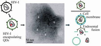
LI Q, LI W, YIN W, et al.. Single-particle tracking of human immunodeficiency virus type 1 productive entry into human primary macrophages[J]. ACS Nano, 2017, 11(4):3890-3903. doi: 10.1021/acsnano.7b00275
|
| [96] |
YU J, ZHANG X J, HAO X J, et al.. Near-infrared fluorescence imaging using organic dye nanoparticles[J]. Biomaterials, 2014, 35(10):3356-3364. doi: 10.1016/j.biomaterials.2014.01.004
|
| [97] |
NI M, ZHUO S, SO P T, et al.. Fluorescent probes for nanoscopy:four categories and multiple possibilities[J]. Journal of Biophotonics, 2017, 10(1):11-23. doi: 10.1002/jbio.201600042
|
| [98] |
NOLLE J M, PRIMPKE S, MULLEN K, et al.. Diffusion of single molecular and macromolecular probes during the free radical bulk polymerization of MMA-towards a better understanding of the Trommsdorff effect on a molecular level[J]. Polym. Chem.-Uk, 2016, 7(24):4100-4105. doi: 10.1039/C6PY00590J
|
| [99] |
HIGGINS D A, PARK S C, TRAN-BA K H, et al.. Single-molecule investigations of morphology and mass transport dynamics in nanostructured materials[J]. Annu. Rev. Anal. Chem., 2015, 8:193-216. doi: 10.1146/annurev-anchem-071114-040153
|
Figures(6)


 DownLoad: