| Citation: | WANG Liang, KONG Wen, HE Yi, HUANG Jiang-jie, SHI Guo-hua. Accurate measurement of mouse eye aberration combined with optical mask modulation[J]. Chinese Optics, 2023, 16(5): 1100-1108. doi: 10.37188/CO.2023-0051

|
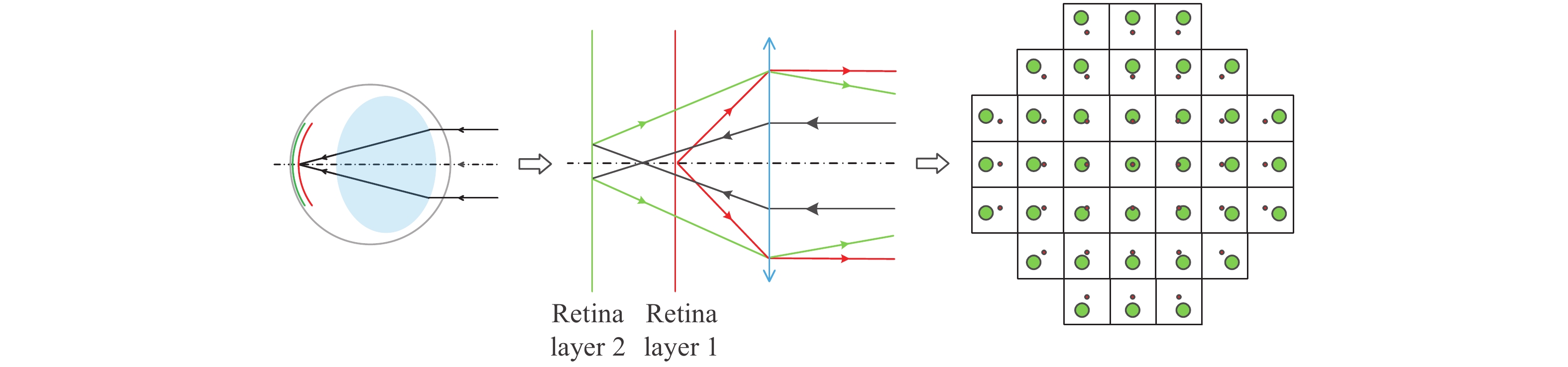
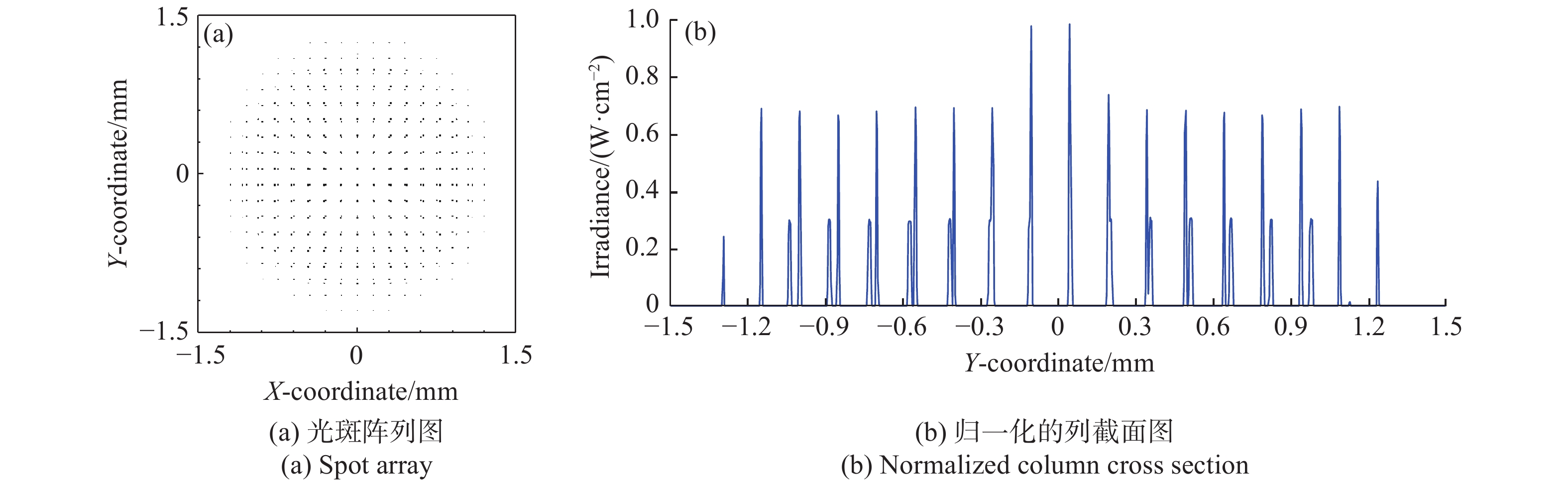
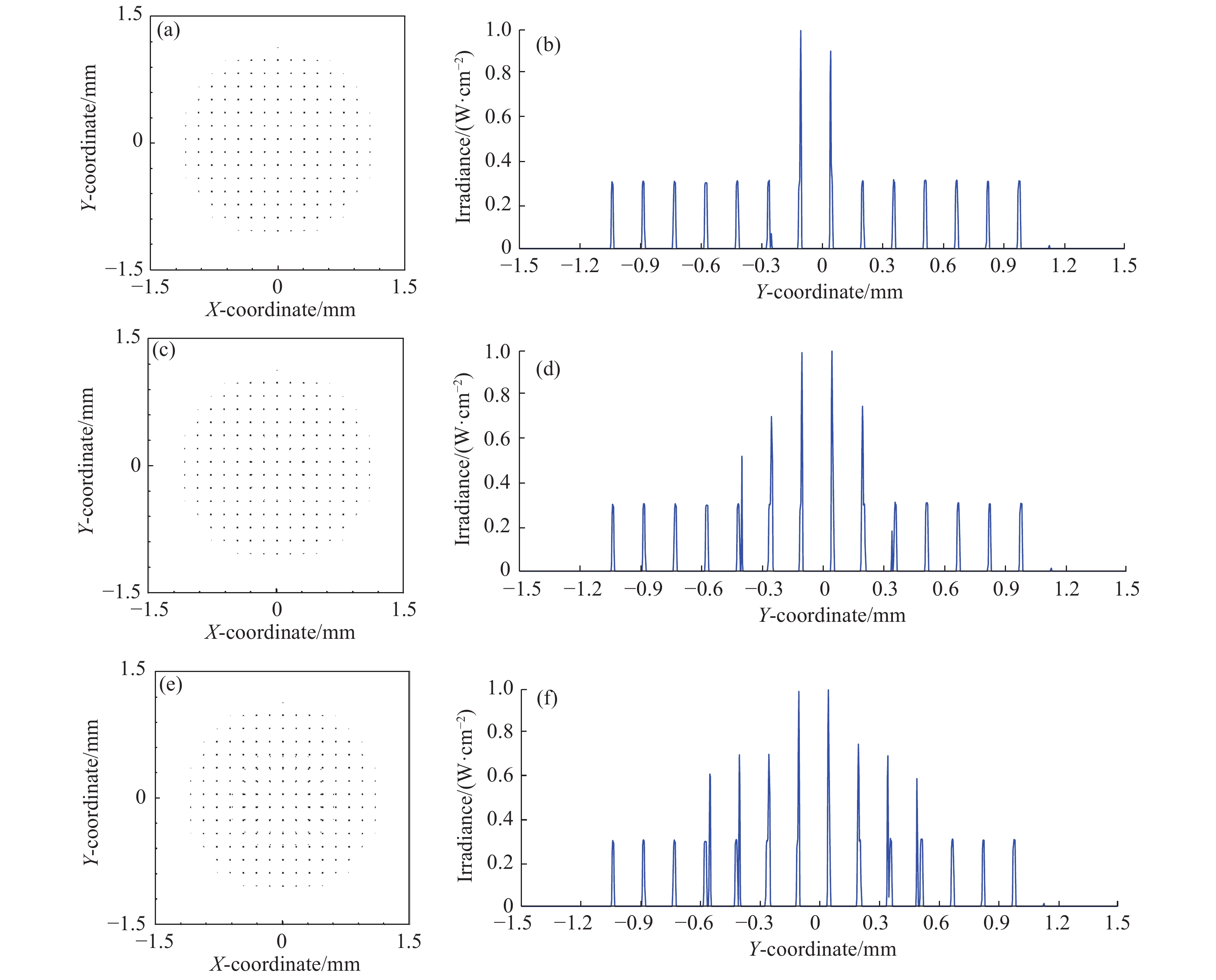
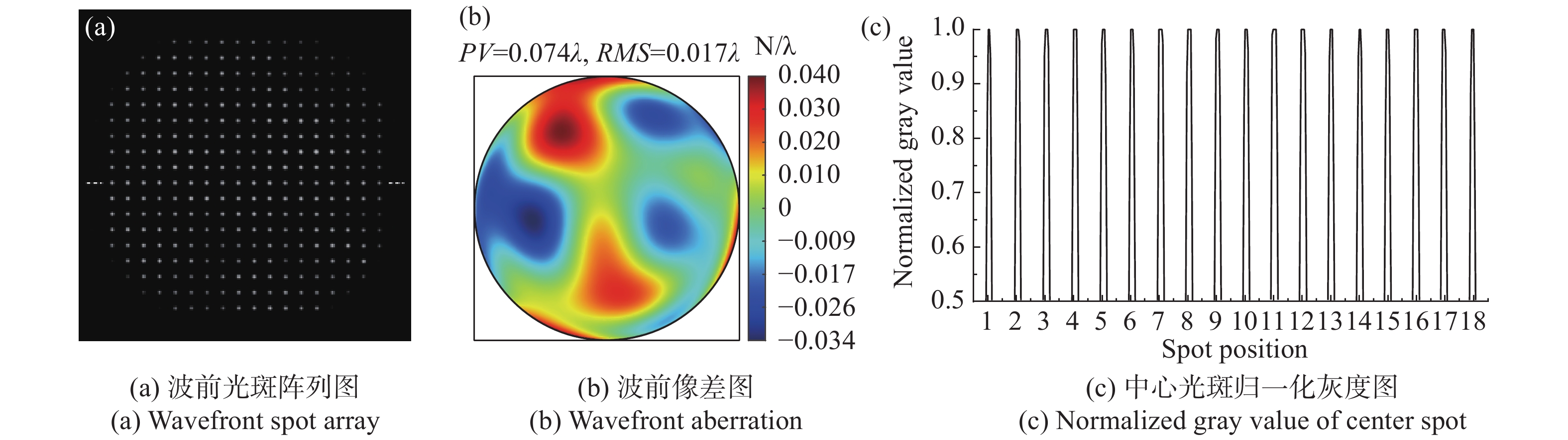
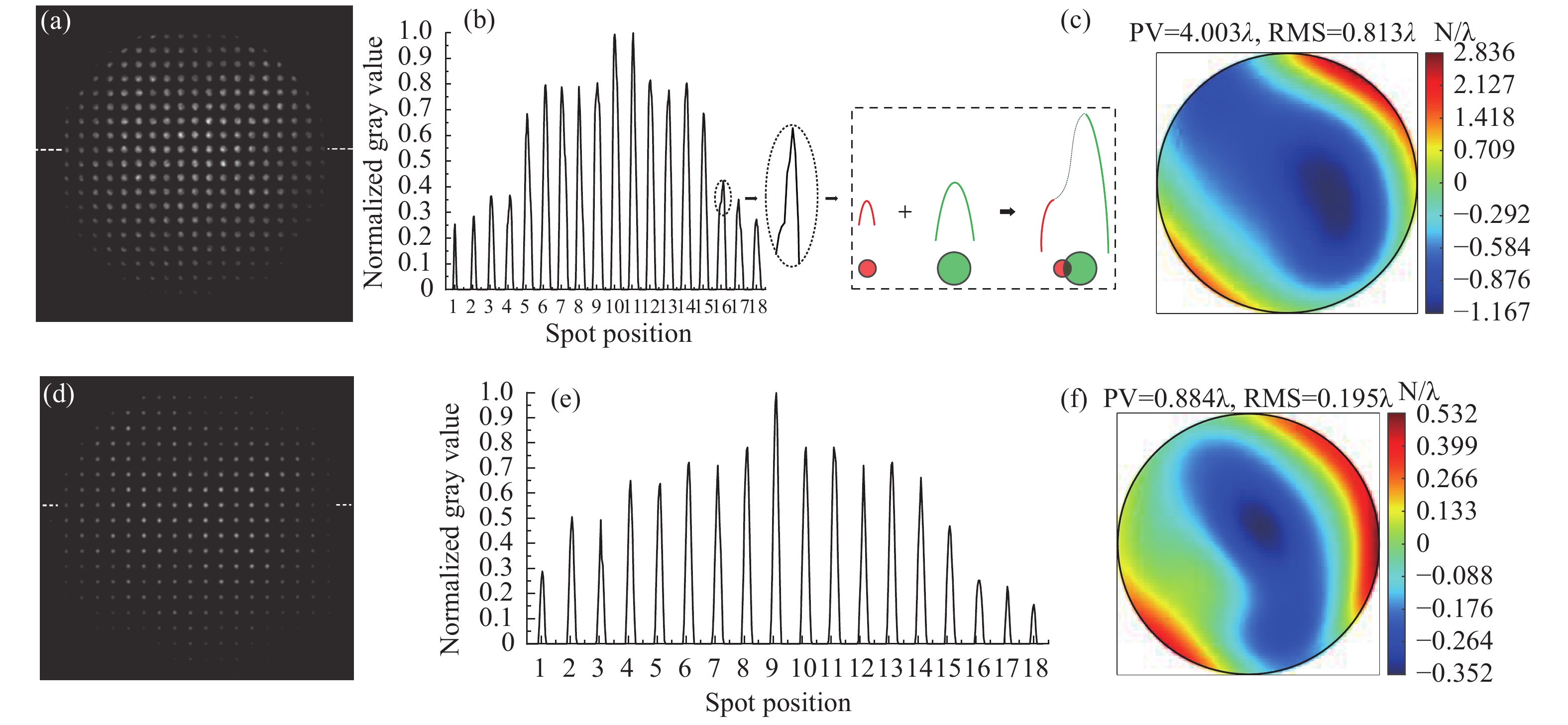
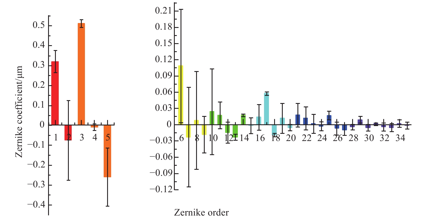
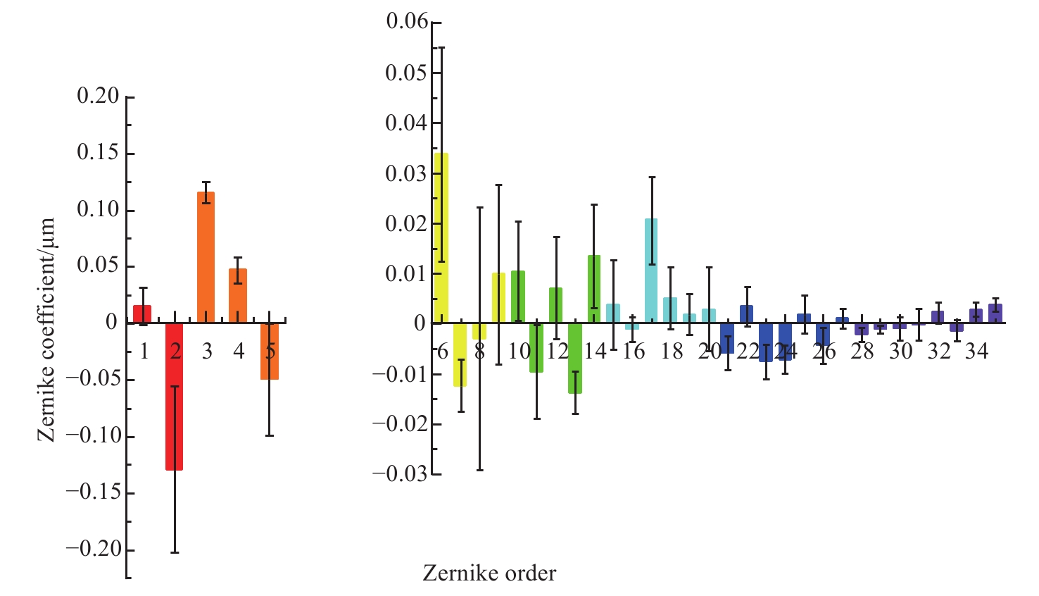
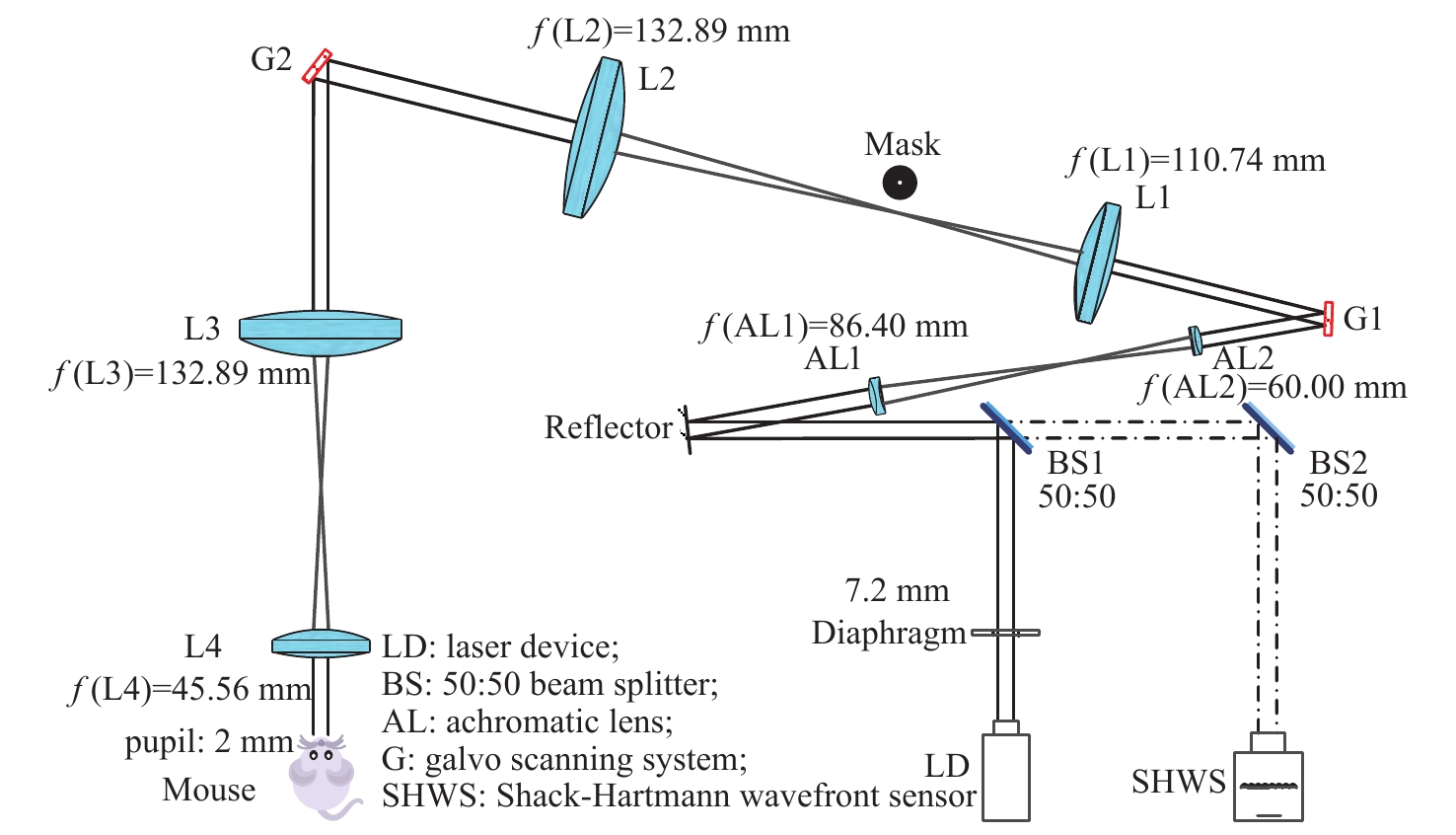
In order to solve the problem of aberration detection failure caused by double-layer reflected light of the fundus retina in standard animal model mouse during wavefront detection, a mouse eye aberration measurement technique combined with optical mask modulation was proposed to improve the accuracy of wavefront aberration measurement. First, according to the key parameters of mouse retina, we established the optical system model of mouse eye wavefront aberration detection and performed optical simulations. Then, the effects of optical masks with different apertures on the reflection beam of the non-target layer of the retina were analyzed and compared, and then the parameters of the optical mask and the experimental plan were determined. Finally, the wave front aberration detection system of the mouse eye was established, and the wavefront aberration of the mouse eye was measured in vivo. The experimental results show that the optical mask with 0.5 mm aperture can reduce the root mean square error of mouse eye wavefront aberration measurement by 74.9%, which is similar to the shielding effect of non-target layer reflected in 80% of the theoretical simulation. It can effectively block the reflected light from the non-target layer of the mouse retina, improve the detection accuracy of the wavefront aberration of the mouse eye, and lay a foundation for the further realization of high-resolution imaging of the mouse eye.

| [1] |
NANEGRUNGSUNK O, PATIKULSILA D, SADDA S R. Ophthalmic imaging in diabetic retinopathy: A review[J]. Clinical &Experimental Ophthalmology, 2022, 50(9): 1082-1096.
|
| [2] |
ROSSI A, RAHIMI M, LE D, et al. Portable widefield fundus camera with high dynamic range imaging capability[J]. Biomedical Optics Express, 2023, 14(2): 906-917. doi: 10.1364/BOE.481096
|
| [3] |
宋宗明, 郭晓红. 眼底多模式影像的进展及其现阶段存在的问题[J]. 中华眼底病杂志,2022,38(2):93-97.
SONG Z M, GUO X H. The progress and problems of the fundus multimodal imaging[J]. Chinese Journal of Ocular Fundus Diseases, 2022, 38(2): 93-97. (in Chinese)
|
| [4] |
唐宁, 樊金宇, 邢利娜, 等. 基于图论的视网膜自动分层方法[J]. 生物医学工程研究,2022,41(2):137-142.
TANG N, FAN J Y, XING L N, et al. Automatic retinal layers segmentation based on graph theory[J]. Journal of Biomedical Engineering Research, 2022, 41(2): 137-142. (in Chinese)
|
| [5] |
MILELLA P, MAPELLI C, NASSISI M, et al. Adaptive optics of kyrieleis plaques in varicella zoster virus-associated posterior uveitis: a multimodal imaging analysis[J]. Journal of Clinical Medicine, 2023, 12(3): 884. doi: 10.3390/jcm12030884
|
| [6] |
GERARDY M, YESILIRMAK N, LEGRAS R, et al. CENTRAL SEROUS CHORIORETINOPATHY: high-resolution imaging of asymptomatic fellow eyes using adaptive optics scanning laser ophthalmoscopy[J]. Retina-the Journal of Retinal and Vitreous Diseases, 2022, 42(2): 375-380.
|
| [7] |
MORGAN J I W, CHUI T Y P, GRIEVE K. Twenty-five years of clinical applications using adaptive optics ophthalmoscopy [Invited][J]. Biomedical Optics Express, 2023, 14(1): 387-428. doi: 10.1364/BOE.472274
|
| [8] |
BISS D P, WEBB R H, ZHOU Y P, et al. An adaptive optics biomicroscope for mouse retinal imaging[J]. Proceedings of SPIE, 2007, 6467: 646703. doi: 10.1117/12.707531
|
| [9] |
张雨东, 姜文汉, 史国华, 等. 自适应光学的眼科学应用[J]. 中国科学 G辑:物理学 力学 天文学,2007,37(1):68-74.
ZHANG Y D, JIANG W H, SHI G H, et al. Application of adaptive optics in ophthalmology[J]. Science in China Physica,Mechanica &Astronomica, 2007, 37(1): 68-74. (in Chinese)
|
| [10] |
LIU L X, WU ZH Q, QI M J, et al. Application of adaptive optics in ophthalmology[J]. Photonics, 2022, 9(5): 288. doi: 10.3390/photonics9050288
|
| [11] |
LIU R X, ZHENG X L, LI D Y, et al. Retinal axial focusing and multi-layer imaging with a liquid crystal adaptive optics camera[J]. Chinese Physics B, 2014, 23(9): 094211. doi: 10.1088/1674-1056/23/9/094211
|
| [12] |
WANG X X, COPMANS D, DE WITTE P A M. Using zebrafish as a disease model to study fibrotic disease[J]. International Journal of Molecular Sciences, 2021, 22(12): 6404. doi: 10.3390/ijms22126404
|
| [13] |
WANG J, CAO H. Zebrafish and medaka: important animal models for human neurodegenerative diseases[J]. International Journal of Molecular Sciences, 2021, 22(19): 10766. doi: 10.3390/ijms221910766
|
| [14] |
YE H, XU X, WANG J X, et al. Polarization effects on the fluorescence emission of zebrafish neurons using light-sheet microscopy[J]. Biomedical Optics Express, 2022, 13(12): 6733-6744. doi: 10.1364/BOE.474588
|
| [15] |
曾雯, 雷玲, 赵铖. 树鼩用于构建自身免疫性疾病动物模型展望[J]. 中国免疫学杂志,2022,38(15):1918-1921.
ZENG W, LEI L, ZHAO CH. Prospects of tree shrews used to establish animal models of autoimmune diseases[J]. Chinese Journal of Immunology, 2022, 38(15): 1918-1921. (in Chinese)
|
| [16] |
JO D H, JANG H K, CHO C S, et al. Visual function restoration in a mouse model of Leber congenital amaurosis via therapeutic base editing[J]. Molecular Therapy-Nucleic Acids, 2023, 31: 16-27. doi: 10.1016/j.omtn.2022.11.021
|
| [17] |
ZHANG M, CHONG K K L, CHEN Z Y, et al. Rapamycin improves Graves' orbitopathy by suppressing CD4+ cytotoxic T lymphocytes[J]. JCI Insight, 2023, 8(3): e160377. doi: 10.1172/jci.insight.160377
|
| [18] |
LI L L, JASMER K J, CAMDEN J M, et al. Early dry eye disease onset in a NOD. H-2h4 mouse model of Sjögren's syndrome[J]. Investigative Ophthalmology &Visual Science, 2022, 63(6): 18.
|
| [19] |
RAMOS R, CABRÉ E, VINYALS A, et al. Orthotopic murine xenograft model of uveal melanoma with spontaneous liver metastasis[J]. Melanoma Research, 2023, 33(1): 1-11. doi: 10.1097/CMR.0000000000000860
|
| [20] |
张鹏飞, 张廷玮, 宋维业, 等. 从小鼠视网膜多种成像方式探讨眼科光学成像技术进展[J]. 中国激光,2020,47(2):0207003. doi: 10.3788/CJL202047.0207003
ZHANG P F, ZHANG T W, SONG W Y, et al. Review of advances in ophthalmic optical imaging technologies from several mouse retinal imaging methods[J]. Chinese Journal of Lasers, 2020, 47(2): 0207003. (in Chinese) doi: 10.3788/CJL202047.0207003
|
| [21] |
GENG Y, DUBRA A, YIN L, et al. Adaptive optics retinal imaging in the living mouse eye[J]. Biomedical Optics Express, 2012, 3(4): 715-734. doi: 10.1364/BOE.3.000715
|
| [22] |
GENG Y, SCHERY L A, SHARMA R, et al. Optical properties of the mouse eye[J]. Biomedical Optics Express, 2011, 2(4): 717-38. doi: 10.1364/BOE.2.000717
|
| [23] |
AKONDI V, DUBRA A. Multi-layer Shack-Hartmann wavefront sensing in the point source regime[J]. Biomedical Optics Express, 2021, 12(1): 409-432. doi: 10.1364/BOE.411189
|
| [24] |
LI Q H, TIMMERS A M, HUNTER K, et al. Noninvasive imaging by optical coherence tomography to monitor retinal degeneration in the mouse[J]. Investigative Ophthalmology &Visual Science, 2001, 42(12): 2981-2989.
|
| [25] |
HORIO N, KACHI S, HORI K, et al. Progressive change of optical coherence tomography scans in retinal degeneration slow mice[J]. Archives of Ophthalmology, 2001, 119(9): 1329-1332. doi: 10.1001/archopht.119.9.1329
|
| [26] |
ABBOTT C J, MCBRIEN N A, GRÜNERT U, et al. Relationship of the optical coherence tomography signal to underlying retinal histology in the tree shrew (Tupaia belangeri)[J]. Investigative Ophthalmology &Visual Science, 2009, 50(1): 414-23.
|
| [27] |
ABBOTT C J, GRÜNERT U, PIANTA M J, et al. Retinal thinning in tree shrews with induced high myopia: Optical coherence tomography and histological assessment[J]. Vision Research, 2011, 51(3): 376-385. doi: 10.1016/j.visres.2010.12.005
|
| [28] |
ZHANG P F, MOCCI J, WAHL D J, et al. Effect of a contact lens on mouse retinal in vivo imaging: Effective focal length changes and monochromatic aberrations[J]. Experimental Eye Research, 2018, 172: 86-93. doi: 10.1016/j.exer.2018.03.027
|
| [29] |
BAWA G, TKATCHENKO T V, AVRUTSKY I, et al. Variational analysis of the mouse and rat eye optical parameters[J]. Biomedical Optics Express, 2013, 4(11): 2585-2595. doi: 10.1364/BOE.4.002585
|
Figures(8) / Tables(1)


 DownLoad: