| Citation: | CHEN Ting-ai, CHEN Long-chao, LI Hui, YU Jia, GAO Yu-feng, ZHENG Wei. Structured illumination super-resolution microscopy technology: review and prospect[J]. Chinese Optics, 2018, 11(3): 307-328. doi: 10.3788/CO.20181103.0307

|

| [1] |
ABBE E. Contributions to the theory of the microscope and that microscopic perception[J]. Arch. Microsc. Anat, 1873, 9:413-468. doi: 10.1007/BF02956173
|
| [2] |
HUANG B, BATES M, ZHUANG X. Super-resolution fluorescence microscopy[J]. Annual Review of Biochemistry, 2009, 78:993-1016. doi: 10.1146/annurev.biochem.77.061906.092014
|
| [3] |
SCHERMELLEH L, HEINTZMANN R, LEONHARDT H. A guide to super-resolution fluorescence microscopy[J]. The Journal of Cell Biology, 2010, 190(2):165-175. doi: 10.1083/jcb.201002018
|
| [4] |
HELL S W, WICHMANN J. Breaking the diffraction resolution limit by stimulated emission:stimulated-emission-depletion fluorescence microscopy[J]. Optics Letters, 1994, 19(11):780-782. doi: 10.1364/OL.19.000780
|
| [5] |
KLAR T A, JAKOBS S, DYBA M, et al.. Fluorescence microscopy with diffraction resolution barrier broken by stimulated emission[J]. Proceedings of the National Academy of Sciences, 2000, 97(15):8206-8210. doi: 10.1073/pnas.97.15.8206
|
| [6] |
BETZIG E, PATTERSON G H, SOUGRAT R, et al.. Imaging intracellular fluorescent proteins at nanometer resolution[J]. Science, 2006, 313(5793):1642-1645. doi: 10.1126/science.1127344
|
| [7] |
HESS S T, GIRIRAJAN T P K, MASON M D. Ultra-high resolution imaging by fluorescence photoactivation localization microscopy[J]. Biophysical Journal, 2006, 91(11):4258-4272. doi: 10.1529/biophysj.106.091116
|
| [8] |
SHROFF H, GALBRAITH C G, GALBRAITH J A, et al.. Live-cell photoactivated localization microscopy of nanoscale adhesion dynamics[J]. Nature Methods, 2008, 5(5):417-423. doi: 10.1038/nmeth.1202
|
| [9] |
RUST M J, BATES M, ZHUANG X. Sub-diffraction-limit imaging by stochastic optical reconstruction microscopy(STORM)[J]. Nature Methods, 2006, 3(10):793-795. doi: 10.1038/nmeth929
|
| [10] |
BATES M, HUANG B, DEMPSEY G T, et al.. Multicolor super-resolution imaging with photo-switchable fluorescent probes[J]. Science, 2007, 317(5845):1749-1753. doi: 10.1126/science.1146598
|
| [11] |
HUANG B, WANG W, BATES M, et al.. Three-dimensional super-resolution imaging by stochastic optical reconstruction microscopy[J]. Science, 2008, 319(5864):810-813. doi: 10.1126/science.1153529
|
| [12] |
HEINTZMANN R, CREMER C. Laterally modulated excitation microscopy:improvement of resolution by using a diffraction grating[J]. Proc. SPIE, 1999, 3568:185-196. doi: 10.1117/12.336833
|
| [13] |
GUSTAFSSON M G L, AGARD D A, SEDAT J W. Doubling the lateral resolution of wide-field fluorescence microscopy using structured illumination[J]. Proc. SPIE, 2000, 3919:141-150. doi: 10.1117/12.384189
|
| [14] |
GUSTAFSSON M G L. Surpassing the lateral resolution limit by a factor of two using structured illumination microscopy[J]. Journal of Microscopy, 2000, 198(2):82-87. doi: 10.1046/j.1365-2818.2000.00710.x
|
| [15] |
WICKER K. Super-resolution fluorescence microscopy using structured illumination[J]. Super-Resolution Microscopy Techniques in the Neurosciences, 2014:133-165. http://cn.bing.com/academic/profile?id=9540a6588212c54c38334ca1d45d2d89&encoded=0&v=paper_preview&mkt=zh-cn
|
| [16] |
REGO E H, SHAO L. Practical structured illumination microscopy[J]. Advanced Fluorescence Microscopy:Methods and Protocols, 2015:175-192. http://cn.bing.com/academic/profile?id=56c13dd845ae9d1fd85b3472f8125184&encoded=0&v=paper_preview&mkt=zh-cn
|
| [17] |
FROHN J T, KNAPP H F, STEMMER A. True optical resolution beyond the Rayleigh limit achieved by standing wave illumination[J]. Proceedings of the National Academy of Sciences, 2000, 97(13):7232-7236. doi: 10.1073/pnas.130181797
|
| [18] |
GUSTAFSSON M G L, SHAO L, CARLTON P M, et al.. Three-dimensional resolution doubling in wide-field fluorescence microscopy by structured illumination[J]. Biophysical Journal, 2008, 94(12):4957-4970. doi: 10.1529/biophysj.107.120345
|
| [19] |
HEINTZMANN R, JOVIN T M, CREMER C. Saturated patterned excitation microscopy-a concept for optical resolution improvement[J]. JOSA A, 2002, 19(8):1599-1609. doi: 10.1364/JOSAA.19.001599
|
| [20] |
GUSTAFSSON M G L. Nonlinear structured-illumination microscopy:wide-field fluorescence imaging with theoretically unlimited resolution[J]. Proceedings of the National Academy of Sciences of the United States of America, 2005, 102(37):13081-13086. doi: 10.1073/pnas.0406877102
|
| [21] |
HIRVONEN L, MANDULA O, WICKER K, et al.. Structured illumination microscopy using photoswitchable fluorescent proteins[J]. Proc. SPIE, 2008, 6861:68610L. doi: 10.1117/12.763021
|
| [22] |
REGO E H, SHAO L, MACKLIN J J, et al.. Nonlinear structured-illumination microscopy with a photoswitchable protein reveals cellular structures at 50-nm resolution[J]. Proceedings of the National Academy of Sciences, 2012, 109(3):E135-E143. doi: 10.1073/pnas.1107547108
|
| [23] |
SCHERMELLEH L, CARLTON P M, HAASE S, et al.. Subdiffraction multicolor imaging of the nuclear periphery with 3D structured illumination microscopy[J]. Science, 2008, 320(5881):1332-1336. doi: 10.1126/science.1156947
|
| [24] |
KNER P, CHHUN B B, GRIFFIS E R, et al.. Super-resolution video microscopy of live cells by structured illumination[J]. Nature Methods, 2009, 6(5):339-342. doi: 10.1038/nmeth.1324
|
| [25] |
SHAO L, KNER P, REGO E H, et al.. Super-resolution 3D microscopy of live whole cells using structured illumination[J]. Nature methods, 2011, 8(12):1044. doi: 10.1038/nmeth.1734
|
| [26] |
LI D, SHAO L, CHEN B C, et al.. Extended-resolution structured illumination imaging of endocytic and cytoskeletal dynamics[J]. Science, 2015, 349(6251):aab3500. doi: 10.1126/science.aab3500
|
| [27] |
CHANG B J, CHOU L J, CHANG Y C, et al.. Isotropic image in structured illumination microscopy patterned with a spatial light modulator[J]. Optics Express, 2009, 17(17):14710-14721. doi: 10.1364/OE.17.014710
|
| [28] |
DAN D, LEI M, YAO B, et al.. DMD-based LED-illumination Super-resolution and optical sectioning microscopy[J]. Scientific Reports, 2013, 3. http://cn.bing.com/academic/profile?id=aed34c513eee106e6e52347ab0ca1b6a&encoded=0&v=paper_preview&mkt=zh-cn
|
| [29] |
LUKEŠ T, KŘÍŽEK P, ŠVINDRYCH Z, et al.. Three-dimensional super-resolution structured illumination microscopy with maximum a posteriori probability image estimation[J]. Optics express, 2014, 22(24):29805-29817. doi: 10.1364/OE.22.029805
|
| [30] |
FÖRSTER R, LU-WALTHER H W, JOST A, et al.. Simple structured illumination microscope setup with high acquisition speed by using a spatial light modulator[J]. Optics Express, 2014, 22(17):20663-20677. doi: 10.1364/OE.22.020663
|
| [31] |
LU-WALTHER H W, KIELHORN M, F RSTER R, et al.. fastSIM:a practical implementation of fast structured illumination microscopy[J]. Methods and Applications in Fluorescence, 2015, 3(1):014001. doi: 10.1088/2050-6120/3/1/014001
|
| [32] |
SONG L, LU-WALTHER H W, F RSTER R, et al.. Fast structured illumination microscopy using rolling shutter cameras[J]. Measurement Science and Technology, 2016, 27(5):055401. doi: 10.1088/0957-0233/27/5/055401
|
| [33] |
YOUNG L J, STR? HL F, KAMINSKI C F. A guide to structured illumination TIRF microscopy at high speed with multiple colors[J]. Journal of Visualized Experiments:JoVE, 2016(111). http://cn.bing.com/academic/profile?id=fa3d794d3a8262b6e8930707da190dd7&encoded=0&v=paper_preview&mkt=zh-cn
|
| [34] |
文刚, 李思黾, 杨西斌, 等.基于激光干涉的结构光照明超分辨荧光显微镜系统[J].光学学报, 2017, 37(3):32-42. http://mall.cnki.net/magazine/Article/GXXB201703004.htm
WEN G, LI S M, YANG X B, et al.. Super-resolution fluorescence microscopy system by structured illumination based on laser interference[J]. Chinese Journal of Optics, 2017, 37(3):32-42.(in Chinese) http://mall.cnki.net/magazine/Article/GXXB201703004.htm
|
| [35] |
SCHAEFER L H, SCHUSTER D, SCHAFFER J. Structured illumination microscopy:artefact analysis and reduction utilizing a parameter optimization approach[J]. Journal of Microscopy, 2004, 216(2):165-174. doi: 10.1111/j.0022-2720.2004.01411.x
|
| [36] |
SHROFF S A, FIENUP J R, WILLIAMS D R. Phase-shift estimation in sinusoidally illuminated images for lateral superresolution[J]. JOSA A, 2009, 26(2):413-424. doi: 10.1364/JOSAA.26.000413
|
| [37] |
SHROFF S A, FIENUP J R, WILLIAMS D R. Lateral superresolution using a posteriori phase shift estimation for a moving object:experimental results[J]. JOSA A, 2010, 27(8):1770-1782. doi: 10.1364/JOSAA.27.001770
|
| [38] |
O'HOLLERAN K, SHAW M. Polarization effects on contrast in structured illumination microscopy[J]. Optics Letters, 2012, 37(22):4603-4605. doi: 10.1364/OL.37.004603
|
| [39] |
WICKER K. Non-iterative determination of pattern phase in structured illumination microscopy using auto-correlations in Fourier space[J]. Optics Express, 2013, 21(21):24692-24701. doi: 10.1364/OE.21.024692
|
| [40] |
WICKER K, MANDULA O, BEST G, et al.. Phase optimisation for structured illumination microscopy[J]. Optics Express, 2013, 21(2):2032-2049. doi: 10.1364/OE.21.002032
|
| [41] |
WICKER K. Non-iterative determination of pattern phase in structured illumination microscopy using auto-correlations in Fourier space[J]. Optics Express, 2013, 21(21):24692-24701. doi: 10.1364/OE.21.024692
|
| [42] |
AYUK R, GIOVANNINI H, JOST A, et al.. Structured illumination fluorescence microscopy with distorted excitations using a filtered blind-SIM algorithm[J]. Optics Letters, 2013, 38(22):4723-4726. doi: 10.1364/OL.38.004723
|
| [43] |
O'HOLLERAN K, SHAW M. Optimized approaches for optical sectioning and resolution enhancement in 2D structured illumination microscopy[J]. Biomedical Optics Express, 2014, 5(8):2580-2590. doi: 10.1364/BOE.5.002580
|
| [44] |
CHU K, MCMILLAN P J, SMITH Z J, et al.. Image reconstruction for structured-illumination microscopy with low signal level[J]. Optics Express, 2014, 22(7):8687-8702. doi: 10.1364/OE.22.008687
|
| [45] |
CHAKROVA N, HEINTZMANN R, RIEGER B, et al.. Studying different illumination patterns for resolution improvement in fluorescence microscopy[J]. Optics Express, 2015, 23(24):31367-31383. doi: 10.1364/OE.23.031367
|
| [46] |
KRÍEK P, LUKEŠ T, OVESNY M, et al.. SIMToolbox:a MATLAB toolbox for structured illumination fluorescence microscopy[J]. Bioinformatics, 2015, 32(2):318-320. http://cn.bing.com/academic/profile?id=d6f0b50129974e99b9099014126be939&encoded=0&v=paper_preview&mkt=zh-cn
|
| [47] |
BALL G, DEMMERLE J, KAUFMANN R, et al.. SIMcheck:a toolbox for successful super-resolution structured illumination microscopy[J]. Scientific Reports, 2015, 5. http://cn.bing.com/academic/profile?id=1f183575f5f5701a11b8b4425ec920a3&encoded=0&v=paper_preview&mkt=zh-cn
|
| [48] |
FÖRSTER R, WICKER K, MVLLER W, et al.. Motion artefact detection in structured illumination microscopy for live cell imaging[J]. Optics Express, 2016, 24(19):22121-22134. doi: 10.1364/OE.24.022121
|
| [49] |
ZHOU X, LEI M, DAN D, et al.. Image recombination transform algorithm for superresolution structured illumination microscopy[J]. Journal of Biomedical Optics, 2016, 21(9):096009-096009. doi: 10.1117/1.JBO.21.9.096009
|
| [50] |
CHAKROVA N, RIEGER B, STALLINGA S. Deconvolution methods for structured illumination microscopy[J]. JOSA A, 2016, 33(7):B12-B20. doi: 10.1364/JOSAA.33.000B12
|
| [51] |
PEREZ V, CHANG B J, STELZER E H K. Optimal 2D-SIM reconstruction by two filtering steps with Richardson-Lucy deconvolution[J]. Scientific Reports, 2016, 6:37149. doi: 10.1038/srep37149
|
| [52] |
YANG Q, CAO L, ZHANG H, et al.. Method of lateral image reconstruction in structured illumination microscopy with super resolution[J]. Journal of Innovative Optical Health Sciences, 2016, 9(3):1630002. doi: 10.1142/S1793545816300020
|
| [53] |
LAL A, SHAN C, XI P. Structured illumination microscopy image reconstruction algorithm[J]. IEEE Journal of Selected Topics in Quantum Electronics, 2016, 22(4):50-63. doi: 10.1109/JSTQE.2016.2521542
|
| [54] |
MVLLER M, MÖNKEMÖLLER V, HENNIG S, et al.. Open-source image reconstruction of super-resolution structured illumination microscopy data in ImageJ[J]. Nature Communications, 2016, 7. http://cn.bing.com/academic/profile?id=94f0b222305508885bd5b06dd8a7c8e4&encoded=0&v=paper_preview&mkt=zh-cn
|
| [55] |
ZHOU X, DAN D, QIAN J, et al.. Super-resolution reconstruction theory in structured illumination microscopy[J]. Chinese Journal of Optics, 2017, 37(3):10-21.(in Chinese) http://cn.bing.com/academic/profile?id=5a55d389721d98c9b9edc18fbe7ac4a2&encoded=0&v=paper_preview&mkt=zh-cn
|
| [56] |
赵天宇, 周兴, 但旦, 等.结构光照明显微中的偏振控制[J].物理学报, 2017, 66(14):295-305. http://www.cnki.com.cn/Article/CJFDTOTAL-JGDJ201501003.htm
ZHAO T Y, ZHOU X, DAN D, et al.. Polarization control methods in structured illumination microscopy[J]. Chinese Journal of Physics, 2017, 66(14):295-305.(in Chinese) http://www.cnki.com.cn/Article/CJFDTOTAL-JGDJ201501003.htm
|
| [57] |
DEMMERLE J, INNOCENT C, NORTH A J, et al.. Strategic and practical guidelines for successful structured illumination microscopy[J]. Nat. Protoc, 2017. http://cn.bing.com/academic/profile?id=a01479c4c15054cf69dd9935ec5bbcbd&encoded=0&v=paper_preview&mkt=zh-cn
|
| [58] |
KRAUS F, MIRON E, DEMMERLE J, et al.. Quantitative 3D structured illumination microscopy of nuclear structures[J]. Nature Protocols, 2017, 12(5):1011-1028. doi: 10.1038/nprot.2017.020
|
| [59] |
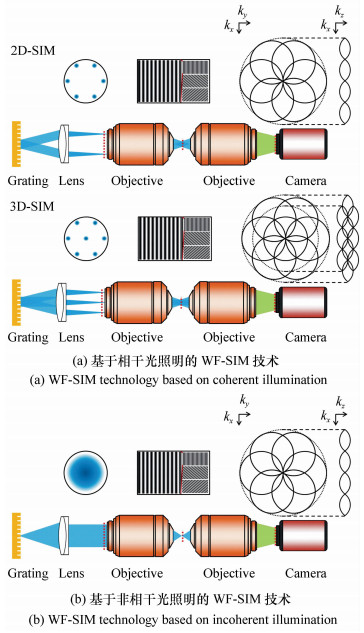
STRÖHL F, KAMINSKI C F. Frontiers in structured illumination microscopy[J]. Optica, 2016, 3(6):667-677. doi: 10.1364/OPTICA.3.000667
|
| [60] |
MVLLER C B, ENDERLEIN J. Image scanning microscopy[J]. Physical Review Letters, 2010, 104(19):198101. doi: 10.1103/PhysRevLett.104.198101
|
| [61] |
YORK A G, PAREKH S H, DALLENOGARE D, et al.. Resolution doubling in live, multicellular organisms via multifocal structured illumination microscopy[J]. Nature Methods, 2012, 9(7):749-754. doi: 10.1038/nmeth.2025
|
| [62] |
SCHULZ O, PIEPER C, CLEVER M, et al.. Resolution doubling in fluorescence microscopy with confocal spinning-disk image scanning microscopy[J]. Proceedings of the National Academy of Sciences, 2013, 110(52):21000-21005. doi: 10.1073/pnas.1315858110
|
| [63] |
SHEPPARD C J R, MEHTA S B, HEINTZMANN R. Superresolution by image scanning microscopy using pixel reassignment[J]. Optics Letters, 2013, 38(15):2889-2892. doi: 10.1364/OL.38.002889
|
| [64] |
DE LUCA G M R, BREEDIJK R M P, BRANDT R A J, et al.. Re-scan confocal microscopy:scanning twice for better resolution[J]. Biomedical Optics Express, 2013, 4(11):2644-2656. doi: 10.1364/BOE.4.002644
|
| [65] |
ROTH S, SHEPPARD C J R, WICKER K, et al.. Optical photon reassignment microscopy(OPRA)[J]. Optical Nanoscopy, 2013, 2(1):5. doi: 10.1186/2192-2853-2-5
|
| [66] |
YORK A G, CHANDRIS P, DALLENOGARE D, et al.. Instant super-resolution imaging in live cells and embryos via analog image processing[J]. Nature Methods, 2013, 10(11):1122-1126. doi: 10.1038/nmeth.2687
|
| [67] |
INGARAMO M, YORK A G, WAWRZUSIN P, et al.. Two-photon excitation improves multifocal structured illumination microscopy in thick scattering tissue[J]. Proceedings of the National Academy of Sciences, 2014, 111(14):5254-5259. doi: 10.1073/pnas.1314447111
|
| [68] |
WINTER P W, YORK A G, DALLENOGARE D, et al.. Two-photon instant structured illumination microscopy improves the depth penetration of super-resolution imaging in thick scattering samples[J]. Optica, 2014, 1(3):181-191. doi: 10.1364/OPTICA.1.000181
|
| [69] |
WEISSHART K. The basic principle of airyscanning[J]. Zeiss Technology Note, 2014:22. http://forum.sci.ccny.cuny.edu/cores/microscopy-imaging/confocal-microscopy/documents/Basic-Principle-Airyscan.pdf
|
| [70] |
STRÖHL F, KAMINSKI C F. A joint Richardson-Lucy deconvolution algorithm for the reconstruction of multifocal structured illumination microscopy data[J]. Methods and Applications in Fluorescence, 2015, 3(1):014002. doi: 10.1088/2050-6120/3/1/014002
|
| [71] |
AZUMA T, KEI T. Super-resolution spinning-disk confocal microscopy using optical photon reassignment[J]. Optics Express, 2015, 23(11):15003-15011. doi: 10.1364/OE.23.015003
|
| [72] |
CURD A, CLEASBY A, MAKOWSKA K, et al.. Construction of an instant structured illumination microscope[J]. Methods, 2015, 88:37-47. doi: 10.1016/j.ymeth.2015.07.012
|
| [73] |
MCGREGOR J E, MITCHELL C A, HARTELL N A. Post-processing strategies in image scanning microscopy[J]. Methods, 2015, 88:28-36. doi: 10.1016/j.ymeth.2015.05.002
|
| [74] |
SIVAGURU M, URBAN M A, FRIED G, et al.. Comparative performance of airyscan and structured illumination superresolution microscopy in the study of the surface texture and 3D shape of pollen[J]. Microscopy Research and Technique, 2016. http://cn.bing.com/academic/profile?id=84817ebfbfa24dc31391778351cd6f06&encoded=0&v=paper_preview&mkt=zh-cn
|
| [75] |
ROTH S, HEINTZMANN R. Optical photon reassignment with increased axial resolution by structured illumination[J]. Methods and Applications in Fluorescence, 2016, 4(4):045005. doi: 10.1088/2050-6120/4/4/045005
|
| [76] |
SHEPPARD C J R, ROTH S, HEINTZMANN R, et al.. Interpretation of the optical transfer function:Significance for image scanning microscopy[J]. Optics Express, 2016, 24(24):27280-27287. doi: 10.1364/OE.24.027280
|
| [77] |
DE LUCA G M R, DESCLOS E, BREEDIJK R M P, et al.. Configurations of the Re-scan Confocal Microscope(RCM) for biomedical applications[J]. Journal of Microscopy, 2017, 266(2):166-177. doi: 10.1111/jmi.2017.266.issue-2
|
| [78] |
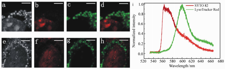
CHEN L C, ZHENG W, et al. . Multicolor re-scan super-resolution imaging of live cells(unpublished).
|
| [79] |
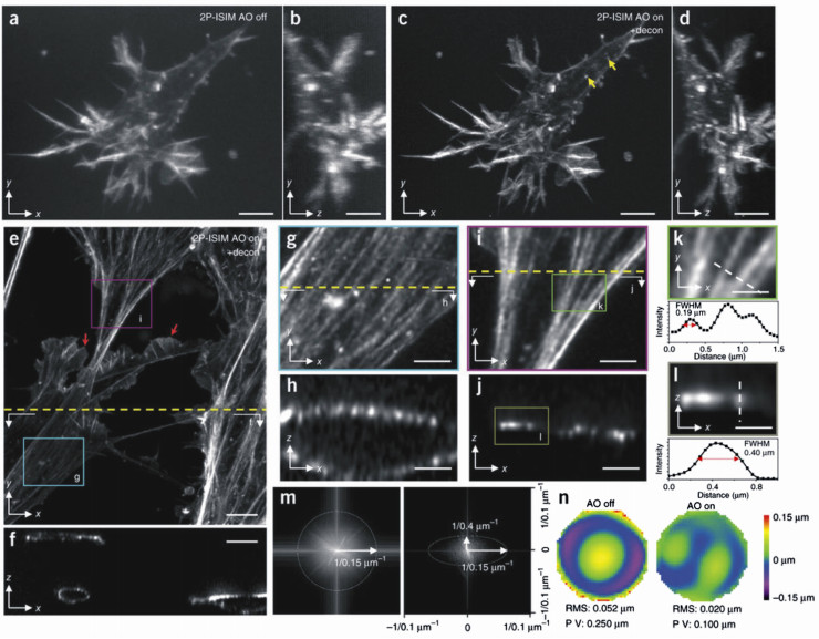
ZHENG W, WU Y, WINTER P, et al.. Adaptive optics improves multiphoton super-resolution imaging[J]. Nature Methods, 2017, 14(9):869-872. doi: 10.1038/nmeth.4337
|
Figures(7) / Tables(2)


 DownLoad: