| Citation: | GUO Hao-hu, GAO Ruo-qian, GE Ming-feng, DONG Wen-fei, LIU Yan, ZHAO Xu-feng. Coronary artery angiography image vessel segmentation method based on feature pyramid network[J]. Chinese Optics, 2024, 17(4): 971-981. doi: 10.37188/CO.2023-0186

|
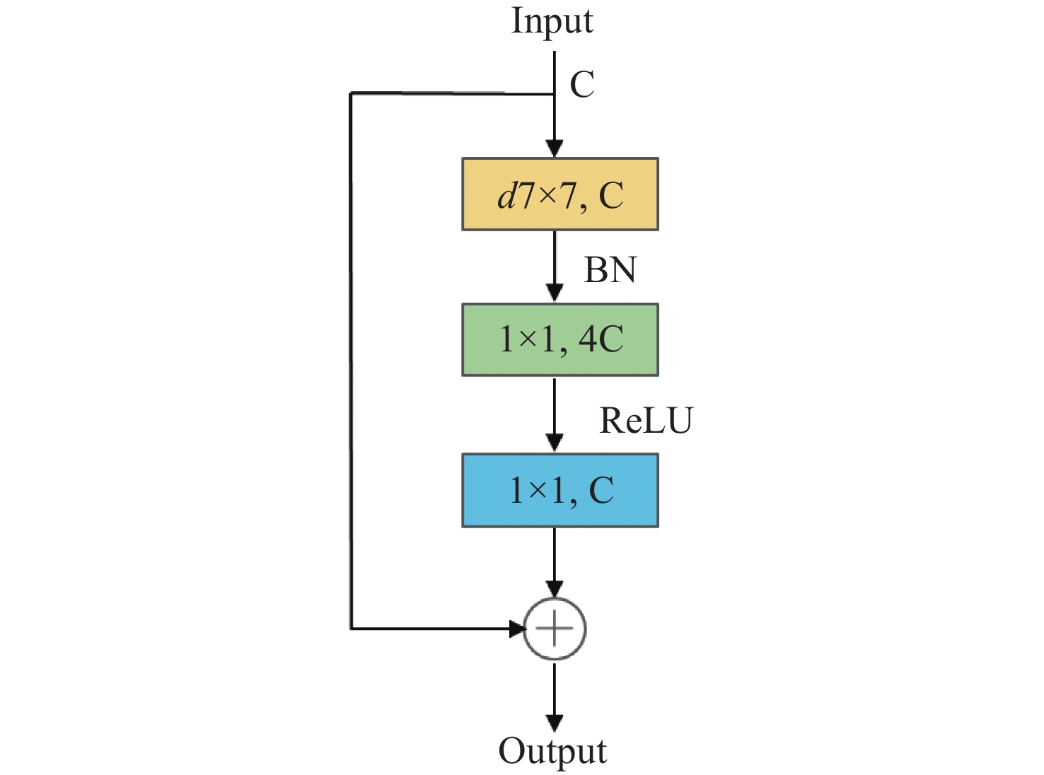
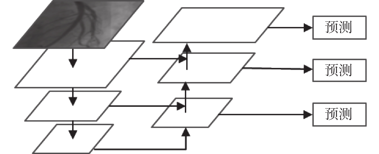
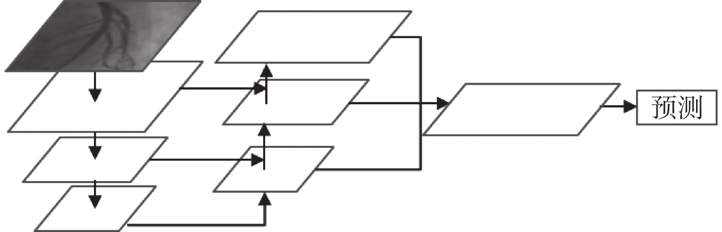
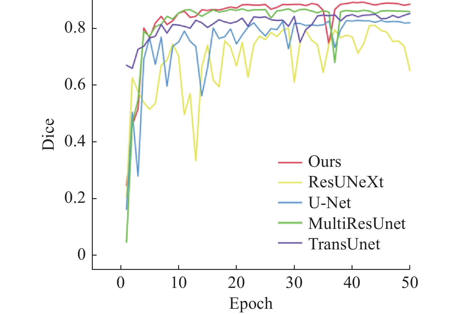
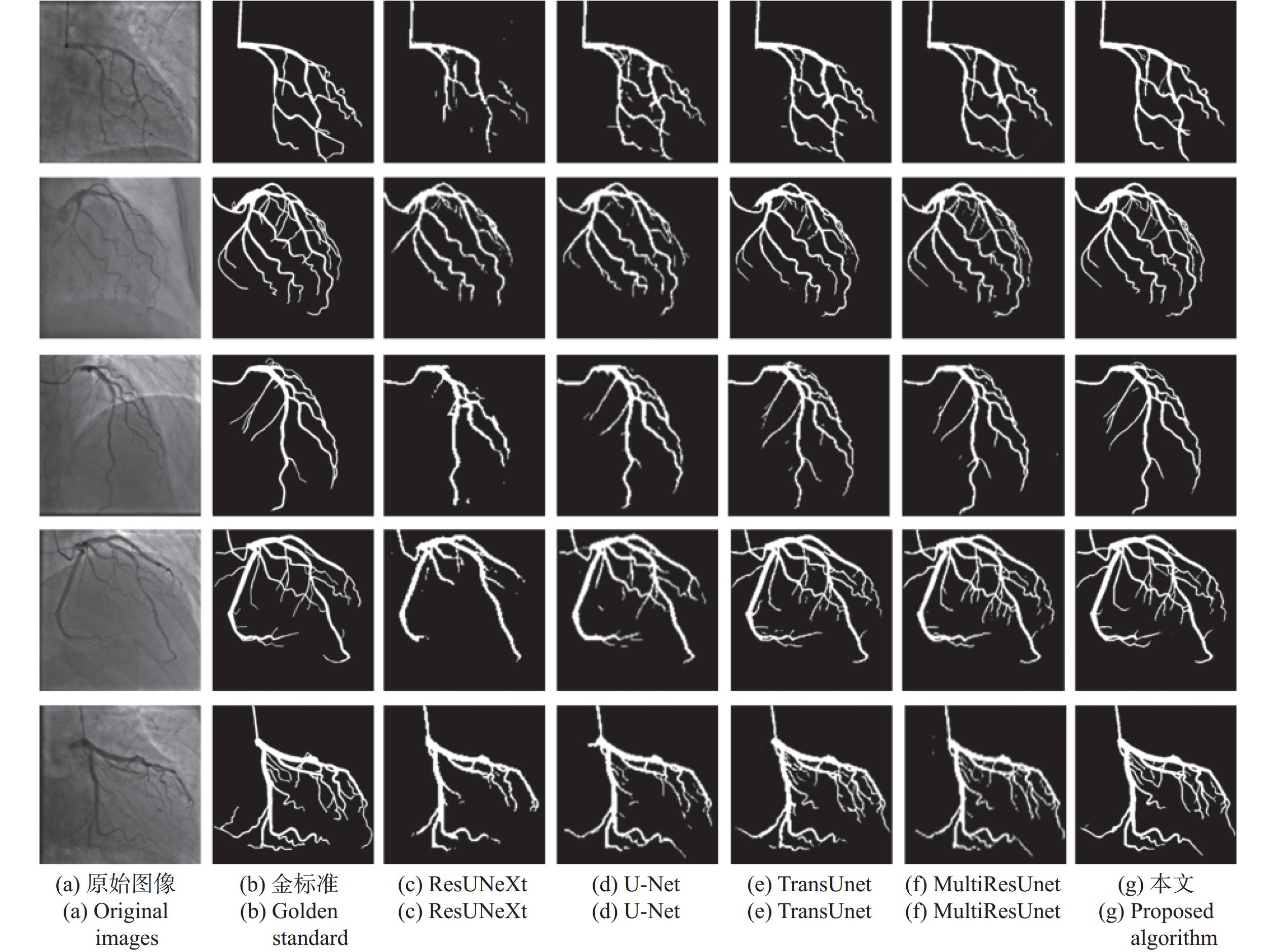
To address issues such as uneven illumination in coronary angiography images, low contrast between vascular structures and background regions, and the complexity of coronary vascular topology, we establish a coronary angiography vascular segmentation annotation dataset. Additionally, we propose a coronary angiography image vascular segmentation model based on the feature map pyramid. On the basis of the U-Net architecture, this model was improved and optimized. First, the first convolutional layer in the U-Net encoding part was replaced with a 7×7 convolutional layer to increase the receptive field of each layer. Modified ConvNeXt blocks were added to the encoding and decoding layers to enhance the network's ability to extract deeper-level features. Second, a Group Attention (GA) mechanism module was designed and incorporated at the U-Net skip connection to strengthen the features extracted from the encoding part, addressing semantic gaps between the encoder and decoder. Finally, a Pyramid Feature Concatenation (PFC) module was designed at the U-Net decoder, which fused features from different scales. Squeeze-and-Excitaton (SE) attention mechanisms were added to each layer of the PFC to filter out effective information from the feature maps. The loss function of the network is weighted based on the outputs of the PFC module at each layer, serving to supervise the feature extraction process across different layers of the network. The test results of this model on the test set are as follows: the Dice coefficient is 0.8843 and the Jaccard coefficient is 0.7926. Experimental results indicate that this model is highly robust in coronary vascular segmentation, more effectively suppressing noise under low contrast and achieving better segmentation results for coronary vessels when compared to other methods.

| [1] |
WANG H, NAGHAVI M, ALLEN C, et al. Global, regional, and national life expectancy, all-cause mortality, and cause-specific mortality for 249 causes of death, 1980-2015: a systematic analysis for the Global Burden of Disease Study 2015[J]. The Lancet, 2016, 388(10053): 1459-1544.
|
| [2] |
JIANGPING S, ZHE Z, WEI W, et al. Assessment of coronary artery stenosis by coronary angiography: a head-to-head comparison with pathological coronary artery anatomy[J]. Circulation: Cardiovascular Interventions, 2013, 6(3): 262-268.
|
| [3] |
FLEMING R M, KIRKEEIDE R L, SMALLING R W, et al. Patterns in visual interpretation of coronary arteriograms as detected by quantitative coronary arteriography[J]. Journal of the American College of Cardiology, 1991, 18(4): 945-951.
|
| [4] |
WIJNS W, SERRUYS P W, REIBER J H, et al. Quantitative angiography of the left anterior descending coronary artery: correlations with pressure gradient and results of exercise thallium scintigraphy[J]. Circulation, 1985, 71(2): 273-279.
|
| [5] |
GARRONE P, BIONDI-ZOCCAI G, SALVETTI I, et al. Quantitative coronary angiography in the current era: principles and applications[J]. Journal of Interventional Cardiology, 2009, 22(6): 527-536.
|
| [6] |
BLONDEL C, MALANDAIN G, VAILLANT R, et al. Reconstruction of coronary arteries from a single rotational X-ray projection sequence[J]. IEEE Transactions on Medical Imaging, 2006, 25(5): 653-663.
|
| [7] |
SHECHTER G, DEVERNAY F, COSTE-MANIÈRE E, et al. Three-dimensional motion tracking of coronary arteries in biplane cineangiograms[J]. IEEE Transactions on Medical Imaging, 2003, 22(4): 493-503.
|
| [8] |
SUN ZH, ZHOU Y. Assessing cardiac dynamics based on X-ray coronary angiograms[J]. J. Multim., 2013, 8(1): 48-55.
|
| [9] |
FELFELIAN B, FAZLALI H R, KARIMI N, et al. Vessel segmentation in low contrast X-ray angiogram images[C]. 2016 IEEE International Conference on Image Processing (ICIP), IEEE, 2016: 375-379.
|
| [10] |
CHEN Y, ZHANG Y D, YANG J, et al. Curve-like structure extraction using minimal path propagation with backtracking[J]. IEEE Transactions on Image Processing, 2016, 25(2): 988-1003.
|
| [11] |
JIN M X, LI R, JIANG J, et al. Extracting contrast-filled vessels in X-ray angiography by graduated RPCA with motion coherency constraint[J]. Pattern Recognition, 2017, 63: 653-666.
|
| [12] |
BANKHEAD P, SCHOLFIELD C N, MCGEOWN J G, et al. Fast retinal vessel detection and measurement using wavelets and edge location refinement[J]. PLoS One, 2012, 7(3): e32435.
|
| [13] |
LI Y L, ZHOU SH J, WU J H, et al. A novel method of vessel segmentation for X-ray coronary angiography images[C]. 2012 Fourth International Conference on Computational and Information Sciences, IEEE, 2012: 468-471.
|
| [14] |
SOARES J V B, LEANDRO J J G, CESAR R M, et al. Retinal vessel segmentation using the 2-D gabor wavelet and supervised classification[J]. IEEE Transactions on Medical Imaging, 2006, 25(9): 1214-1222.
|
| [15] |
FRANGI A F, NIESSEN W J, VINCKEN K L, et al. Multiscale vessel enhancement filtering[C]. Proceedings of the 1st International Conference on Medical Image Computing and Computer-Assisted Intervention— MICCAI’98, Springer, 1998: 130-137.
|
| [16] |
M’HIRI F, DUONG L, DESROSIERS C, et al. Vessel walker: coronary arteries segmentation using random walks and hessian-based vesselness filter[C]. 2013 IEEE 10th International Symposium on Biomedical Imaging, IEEE, 2013: 918-921.
|
| [17] |
DEHKORDI M T, HOSEINI A M D, SADRI S, et al. Local feature fitting active contour for segmenting vessels in angiograms[J]. IET Computer Vision, 2014, 8(3): 161-170.
|
| [18] |
LAW M W K, CHUNG A C S. Efficient implementation for spherical flux computation and its application to vascular segmentation[J]. IEEE Transactions on Image Processing, 2009, 18(3): 596-612.
|
| [19] |
ORLANDO J I, PROKOFYEVA E, BLASCHKO M B. A discriminatively trained fully connected conditional random field model for blood vessel segmentation in fundus images[J]. IEEE Transactions on Bio-Medical Engineering, 2017, 64(1): 16-27.
|
| [20] |
郑跃坤, 葛明锋, 常智敏, 等. 基于残差网络的结直肠内窥镜图像超分辨率重建方法[J]. 中国光学(中英文),2023,16(5):1022-1033.
ZHENG Y K, GE M F, CHANG ZH M, et al. Super-resolution reconstruction for colorectal endoscopic images based on a residual network[J]. Chinese Optics, 2023, 16(5): 1022-1033. (in Chinese).
|
| [21] |
白瑞峰, 江山, 孙海江, 等. 基于编码解码结构的微血管减压图像实时语义分割[J]. 中国光学(中英文),2022,15(5):1055-1065.
BAI R F, JIANG SH, SUN H J, et al. Real-time semantic segmentation of microvascular decompression images based on encoder-decoder structure[J]. Chinese Optics, 2022, 15(5): 1055-1065. (in Chinese).
|
| [22] |
ZHAO H SH, SHI J P, QI X J, et al. Pyramid scene parsing network[C]. 2017 IEEE Conference on Computer Vision and Pattern Recognition (CVPR), IEEE, 2017: 6230-6239.
|
| [23] |
BADRINARAYANAN V, KENDALL A, CIPOLLA R. SegNet: a deep convolutional encoder-decoder architecture for image segmentation[J]. IEEE Transactions on Pattern Analysis and Machine Intelligence, 2017, 39(12): 2481-2495.
|
| [24] |
RONNEBERGER O, FISCHER P, BROX T. U-Net: convolutional networks for biomedical image segmentation[C]. Proceedings of the 18th International Conference on Medical Image Computing and Computer-Assisted Intervention – MICCAI 2015, Springer, 2015: 234-241.
|
| [25] |
IBTEHAZ N, RAHMAN M S. MultiResUNet: rethinking the U-Net architecture for multimodal biomedical image segmentation[J]. Neural Networks, 2020, 121: 74-87.
|
| [26] |
PAN S W, ZHANG W, ZHANG W J, et al. Diagnostic model of coronary microvascular disease combined with full convolution deep network with balanced cross-entropy cost function[J]. IEEE Access, 2019, 7: 177997-178006.
|
| [27] |
XIAN ZH CH, WANG X Q, YAN SH D, et al. Main coronary vessel segmentation using deep learning in smart medical[J]. Mathematical Problems in Engineering, 2020, 2020: 8858344.
|
| [28] |
YANG S, KWEON J, KIM Y H. Major vessel segmentation on X-ray coronary angiography using deep networks with a novel penalty loss function[C]. Proceedings of Machine Learning Research, MIDL, 2019.
|
| [29] |
JUN T J, KWEON J, KIM Y H, et al. T-Net: Nested encoder-decoder architecture for the main vessel segmentation in coronary angiography[J]. Neural Networks, 2020, 128: 216-233.
|
| [30] |
LI L ZH, VERMA M, NAKASHIMA Y, et al. IterNet: retinal image segmentation utilizing structural redundancy in vessel networks[C]. 2020 IEEE Winter Conference on Applications of Computer Vision (WACV), IEEE, 2020: 3645-3654.
|
| [31] |
LIU ZH, MAO H Z, WU CH Y, et al. A ConvNet for the 2020s[C]. IEEE/CVF Conference on Computer Vision and Pattern Recognition, IEEE, 2022: 11966-11976.
|
| [32] |
LIN T Y, DOLLÁR P, GIRSHICK R, et al. Feature pyramid networks for object detection[C]. 2017 IEEE Conference on Computer Vision and Pattern Recognition (CVPR), IEEE, 2017: 936-944.
|
| [33] |
HU J, SHEN L, SUN G. Squeeze-and-excitation networks[C]. 2018 IEEE/CVF Conference on Computer Vision and Pattern Recognition, IEEE, 2018: 7132-7141.
|
| [34] |
VASWANI A, SHAZEER N, PARMAR N, et al. Attention is all you need[C]. Proceedings of the 31st International Conference on Neural Information Processing Systems, NIPS, 2017: 5998-6008.
|
| [35] |
DIAKOGIANNIS F I, WALDNER F, CACCETTA P, et al. ResUNet-a: a deep learning framework for semantic segmentation of remotely sensed data[J]. ISPRS Journal of Photogrammetry and Remote Sensing, 2020, 162: 94-114.
|
| [36] |
CHEN J N, LU Y Y, YU Q H, et al. TransUNet: transformers make strong encoders for medical image segmentation[J]. arXiv: 2102.04306, 2021.
|

Figures(9) / Tables(2)


 DownLoad: