| Citation: | TAN Tian, SHI Tian-yue, WU Chang-feng, PENG Hong-shang. NIR-II fluorescence confocal imaging based on indirect wavefront shaping[J]. Chinese Optics, 2024, 17(1): 150-159. doi: 10.37188/CO.2023-0070

|
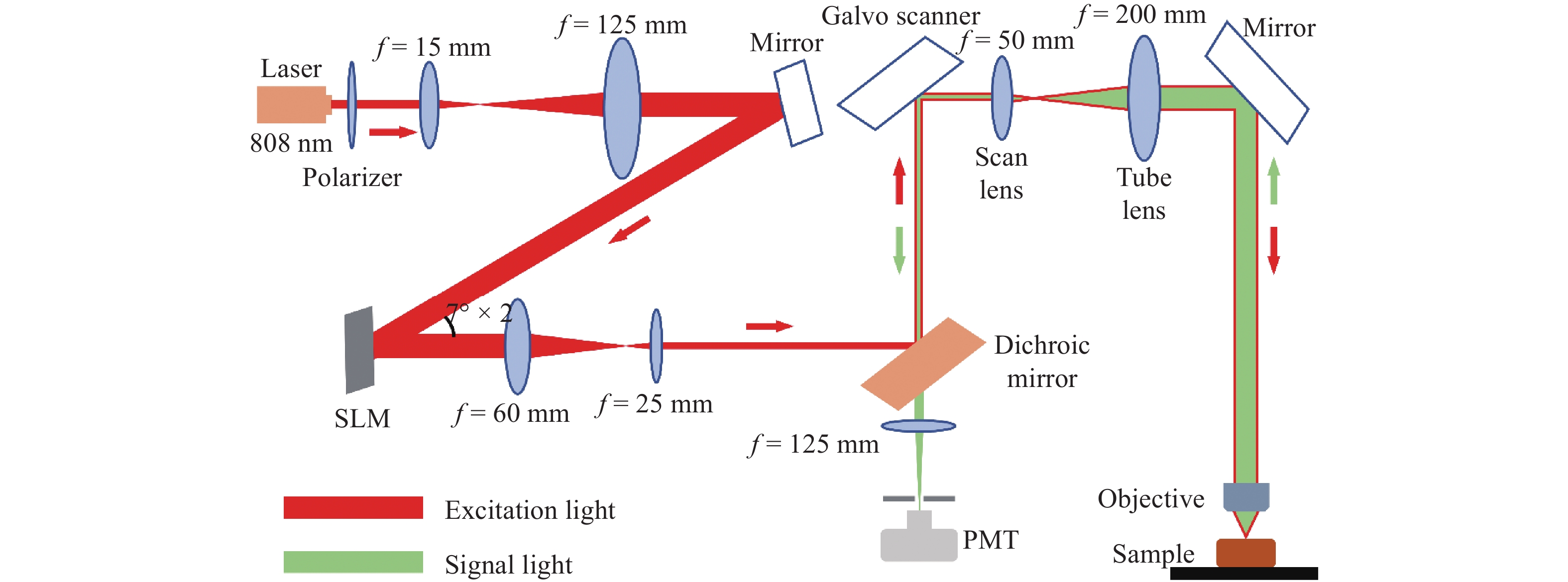
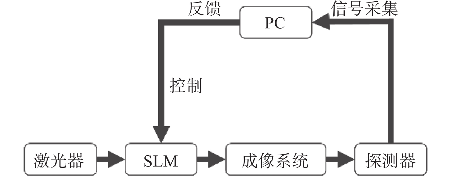
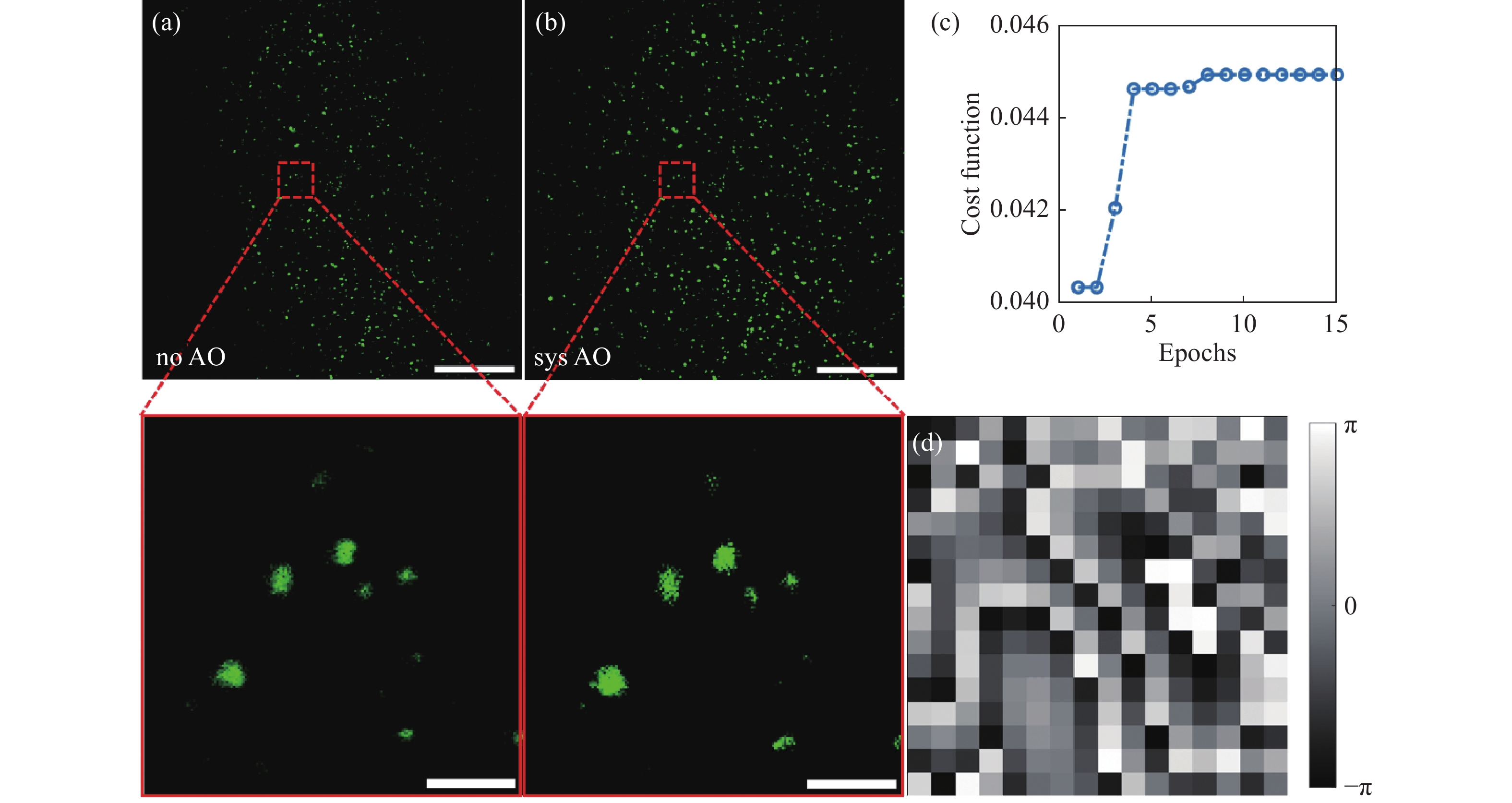
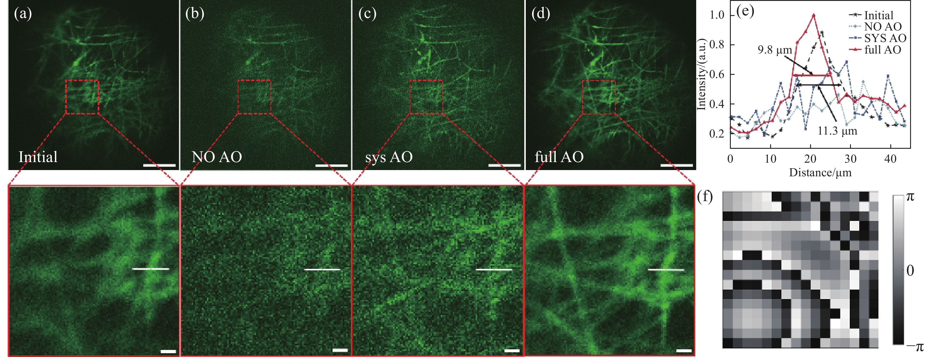
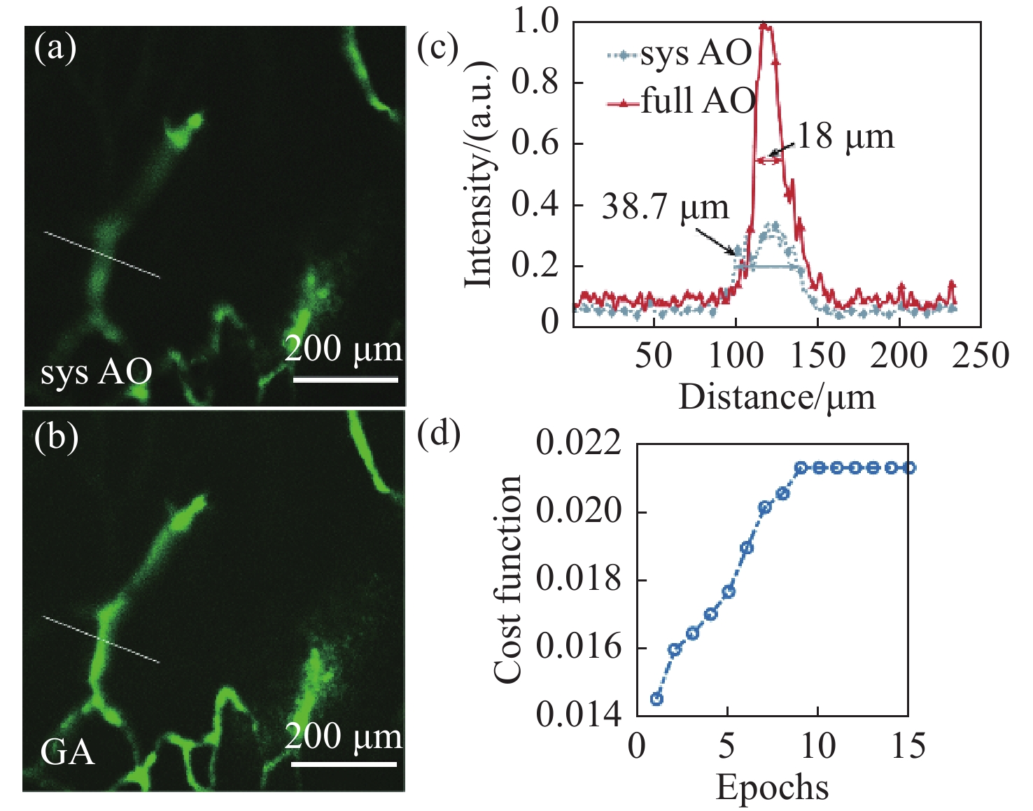
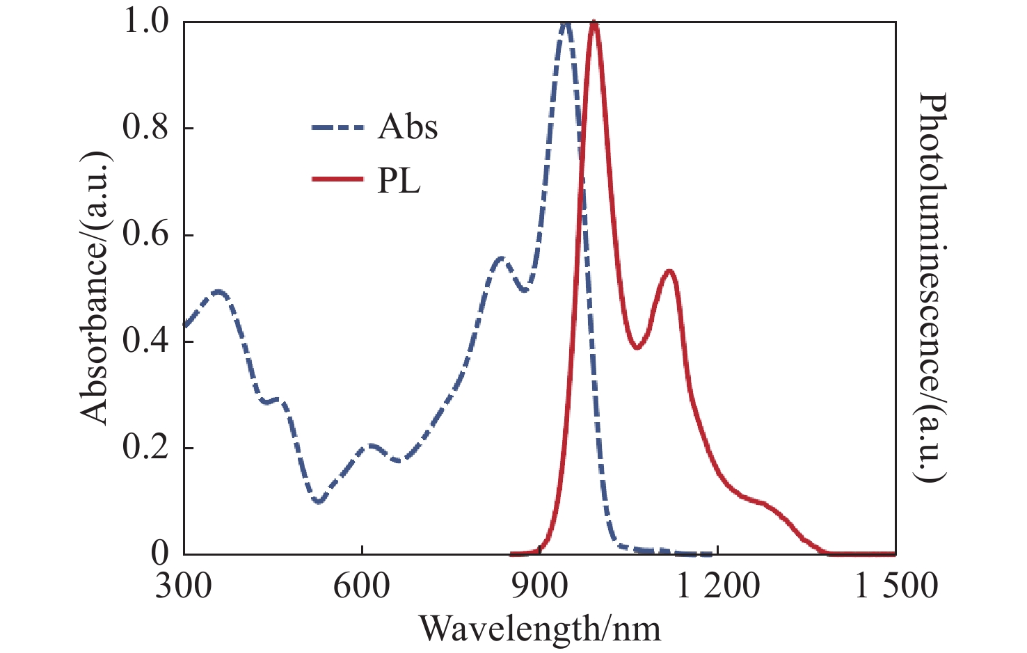
Optical aberrations caused by the scattering of biological tissues limit the imaging performance of optical systems. A near-infrared II fluorescence confocal imaging technique based on indirect wavefront shaping was investigated. First, we synthesized a highly efficient near-infrared II range fluorescent probe, where reducing the scattering of biological tissue can realize biopsy imaging with high-contrast. Second, we investigated the adaptive optical method based on indirect wavefront measurement. The indirect wavefront shaping technology was applied to the laser scanning confocal system, enabling the measurement and compensation of optical aberrations caused by biological tissues, and obtaining imaging of biological tissues with a high signal-to-noise ratio. Finally, near-infrared II fluorescence confocal imaging system based on indirect wavefront shaping was deployed and relevant experiments were conducted. The experimental results indicate that the system effectively compensates for the aberrations induced by air plates, scattering media and mouse skull, and increases the final signal intensity by 1.47, 1.95 and 2.85 times, respectively. As a result, the final imaging quality is significantly enhanced.

| [1] |
KLIMAS A, ZHAO Y X. Expansion microscopy: toward nanoscale imaging of a diverse range of biomolecules[J]. ACS Nano, 2020, 14(7): 7689-7695. doi: 10.1021/acsnano.0c04374
|
| [2] |
ADESNIK H, ABDELADIM L. Probing neural codes with two-photon holographic optogenetics[J]. Nature Neuroscience, 2021, 24(10): 1356-1366. doi: 10.1038/s41593-021-00902-9
|
| [3] |
WEIGELIN B, BAKKER G J, FRIEDL P. Third harmonic generation microscopy of cells and tissue organization[J]. Journal of Cell Science, 2016, 129(2): 245-255.
|
| [4] |
HAMPSON K M, TURCOTTE R, MILLER D T, et al. Adaptive optics for high-resolution imaging[J]. Nature Reviews Methods Primers, 2021, 1(1): 68. doi: 10.1038/s43586-021-00066-7
|
| [5] |
JI N. Adaptive optical fluorescence microscopy[J]. Nature Methods, 2017, 14(4): 374-380. doi: 10.1038/nmeth.4218
|
| [6] |
LIU P F, ZHAO R, LI H W, et al. Near-infrared-II deep tissue fluorescence microscopy and application[J]. Nano Research, 2023, 16(1): 692-714. doi: 10.1007/s12274-022-4836-y
|
| [7] |
杜婉晴, 宋文琦, 梁天宇, 等. 一种基于二氰基异佛尔酮的近红外汞离子荧光探针的合成与应用[J]. 分析化学,2023,51(3):421-428. doi: 10.19756/j.issn.0253-3820.221583
DU W Q, SONG W Q, LIANG T Y, et al. Synthesis and application of near infrared mercury (II) fluorescent probe based on dicyanoisophorone[J]. Chinese Journal of Analytical Chemistry, 2023, 51(3): 421-428. (in Chinese) doi: 10.19756/j.issn.0253-3820.221583
|
| [8] |
张松涛, 王樱蕙, 张洪杰. Nd3+离子敏化的荧光纳米探针用于近红外二区血管成像[J]. 应用化学,2022,39(4):685-693.
ZHANG S T, WANG Y H, ZHANG H J. Nd3+ sensitized fluorescent nanoprobes for vascular imaging in the second near infrared window[J]. Chinese Journal of Applied Chemistry, 2022, 39(4): 685-693. (in Chinese)
|
| [9] |
WELSHER K, SHERLOCK S P, DAI H J. Deep-tissue anatomical imaging of mice using carbon nanotube fluorophores in the second near-infrared window[J]. Proceedings of the National Academy of Sciences of the United States of America, 2011, 108(22): 8943-8948.
|
| [10] |
HONG G S, ZOU Y P, ANTARIS A L, et al. Ultrafast fluorescence imaging in vivo with conjugated polymer fluorophores in the second near-infrared window[J]. Nature Communications, 2014, 5(1): 4206. doi: 10.1038/ncomms5206
|
| [11] |
WANG F F, WAN H, MA Z R, et al. Light-sheet microscopy in the near-infrared II window[J]. Nature Methods, 2019, 16(6): 545-552. doi: 10.1038/s41592-019-0398-7
|
| [12] |
HONG G S, DIAO S, CHANG J L, et al. Through-skull fluorescence imaging of the brain in a new near-infrared window[J]. Nature Photonics, 2014, 8(9): 723-730. doi: 10.1038/nphoton.2014.166
|
| [13] |
桑若愚, 许兴鹏, 王其, 等. 近红外二区有机小分子荧光探针[J]. 化学学报,2020,78(9):901-915. doi: 10.6023/A20050190
SANG R Y, XU X P, WANG Q, et al. Near-infrared-II fluorescence probes based on organic small molecules[J]. Acta Chimica Sinica, 2020, 78(9): 901-915. (in Chinese) doi: 10.6023/A20050190
|
| [14] |
张天宇, 王钢, 张熙, 等. 基于焦面复制方法的自适应光学系统静态像差校正技术[J]. 中国光学,2022,15(3):545-551.
ZHANG T Y, WANG G, ZHANG X, et al. Staticaberration correction technique for adaptive optics system based on focal-plane copy approach[J]. Chinese Optics, 2022, 15(3): 545-551. (in Chinese)
|
| [15] |
YANG J M, HE Q Z, LIU L X, et al. Anti-scattering light focusing by fast wavefront shaping based on multi-pixel encoded digital-micromirror device[J]. Light:Science & Applications, 2021, 10(1): 149.
|
| [16] |
YEMINY T, KATZ O. Guidestar-free image-guided wavefront shaping[J]. Science Advances, 2021, 7(21): eabf5364. doi: 10.1126/sciadv.abf5364
|
| [17] |
JI N, MILKIE D E, BETZIG E. Adaptive optics via pupil segmentation for high-resolution imaging in biological tissues[J]. Nature Methods, 2010, 7(2): 141-147. doi: 10.1038/nmeth.1411
|
| [18] |
AZUCENA O, CREST J, CAO J, et al. Wavefront aberration measurements and corrections through thick tissue using fluorescent microsphere reference beacons[J]. Optics Express, 2010, 18(16): 17521-17532. doi: 10.1364/OE.18.017521
|
| [19] |
TAO X D, FERNANDEZ B, AZUCENA O, et al. Adaptive optics confocal microscopy using direct wavefront sensing[J]. Optics Letters, 2011, 36(7): 1062-1064. doi: 10.1364/OL.36.001062
|
| [20] |
KATZ O, RAMAZ F, GIGAN S, et al. Controlling light in complex media beyond the acoustic diffraction-limit using the acousto-optic transmission matrix[J]. Nature Communications, 2019, 10(1): 717. doi: 10.1038/s41467-019-08583-6
|
| [21] |
ZHOU ZH, HUANG J F, LI X, et al. Adaptive optical microscopy via virtual-imaging-assisted wavefront sensing for high-resolution tissue imaging[J]. PhotoniX, 2022, 3(1): 13. doi: 10.1186/s43074-022-00060-6
|
| [22] |
JI N, SATO T R, BETZIG E. Characterization and adaptive optical correction of aberrations during in vivo imaging in the mouse cortex[J]. Proceedings of the National Academy of Sciences of the United States of America, 2012, 109(1): 22-27.
|
| [23] |
TANG J, GERMAIN R N, CUI M. Superpenetration optical microscopy by iterative multiphoton adaptive compensation technique[J]. Proceedings of the National Academy of Sciences of the United States of America, 2012, 109(22): 8434-8439.
|
| [24] |
PARK J H, SUN W, CUI M. High-resolution in vivo imaging of mouse brain through the intact skull[J]. Proceedings of the National Academy of Sciences of the United States of America, 2015, 112(30): 9236-9241.
|
| [25] |
PAPADOPOULOS I N, JOUHANNEAU J S, POULET J F A, et al. Scattering compensation by focus scanning holographic aberration probing (F-SHARP)[J]. Nature Photonics, 2017, 11(2): 116-123. doi: 10.1038/nphoton.2016.252
|
| [26] |
POZZI P, GANDOLFI D, PORRO C A, et al. Scattering compensation for deep brain microscopy: The long road to get proper images[J]. Frontiers in Physics, 2020, 8: 26. doi: 10.3389/fphy.2020.00026
|
| [27] |
LI R Z, PENG T, LIANG Y SH, et al. Interleaved segment correction achieves higher improvement factors in using genetic algorithm to optimize light focusing through scattering media[J]. Journal of Optics, 2017, 19(10): 105602. doi: 10.1088/2040-8986/aa84dc
|
| [28] |
MAY M A, BARRE N, KUMMER K K, et al. Fast holographic scattering compensation for deep tissue biological imaging[J]. Nature Communications, 2021, 12(1): 4340. doi: 10.1038/s41467-021-24666-9
|
| [29] |
LIU Y, LIU J F, CHEN D D, et al. Fluorination enhances NIR‐II fluorescence of polymer dots for quantitative brain tumor imaging[J]. Angewandte Chemie, 2020, 132(47): 21235-21243. doi: 10.1002/ange.202007886
|
Figures(7)


 DownLoad: