| Citation: | BAI Rui-feng, JIANG Shan, SUN Hai-jiang, LIU Xin-rui. Real-time semantic segmentation of microvascular decompression images based on encoder-decoder structure[J]. Chinese Optics, 2022, 15(5): 1055-1065. doi: 10.37188/CO.2022-0120

|
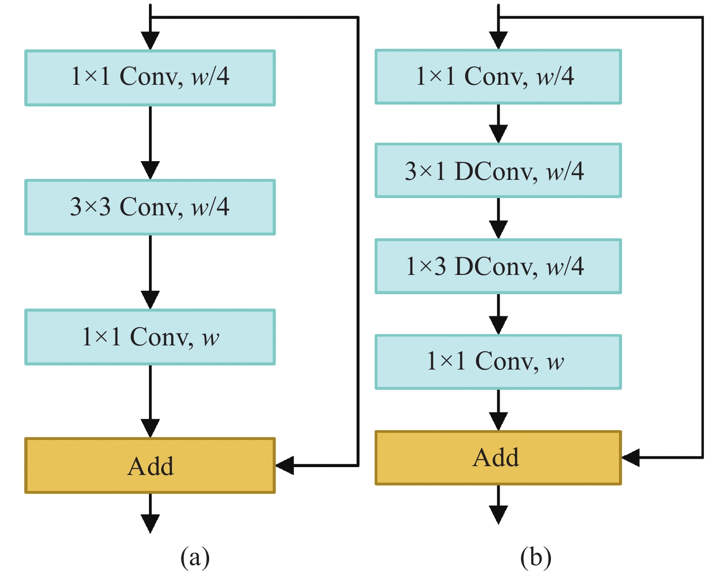
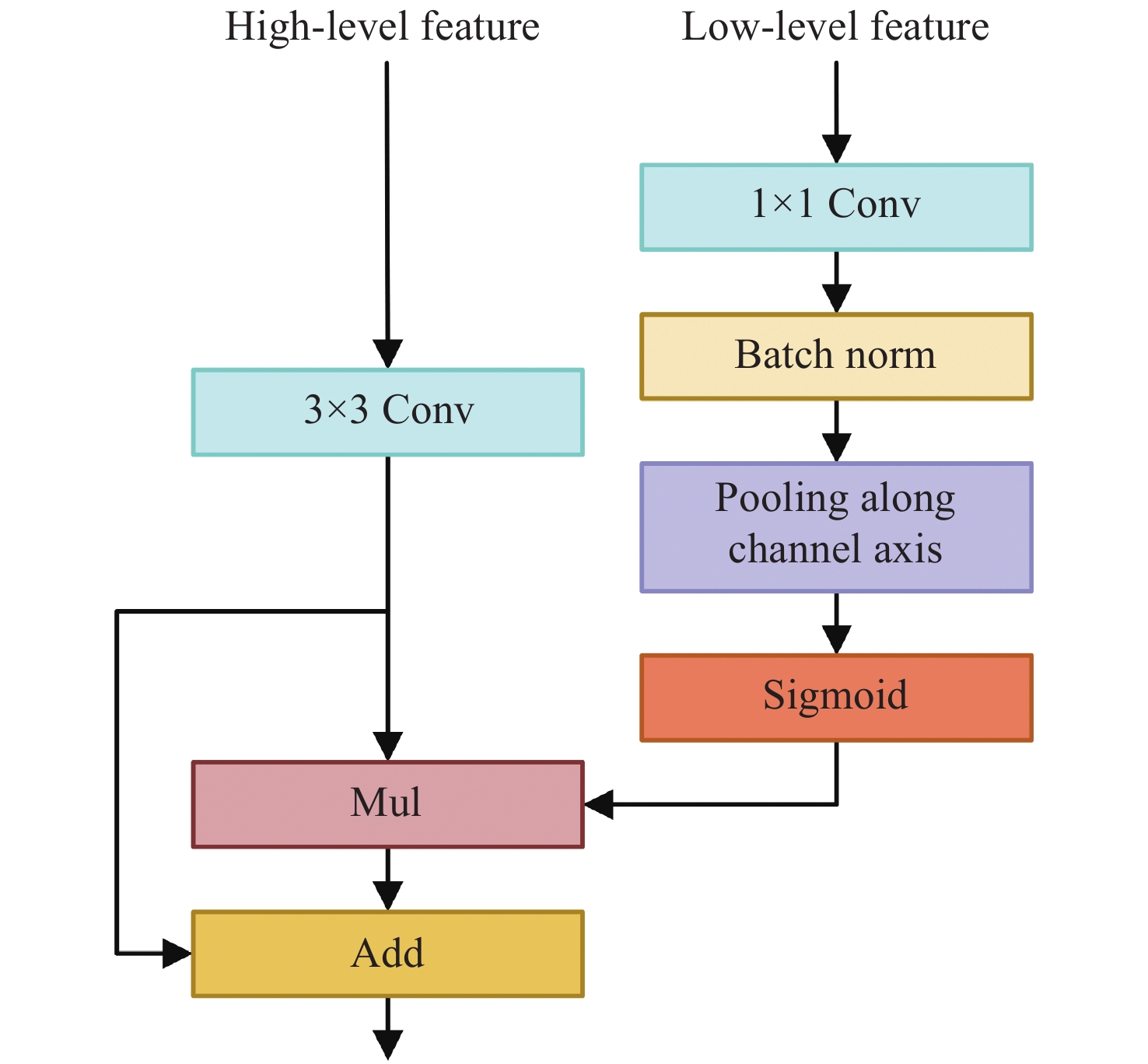
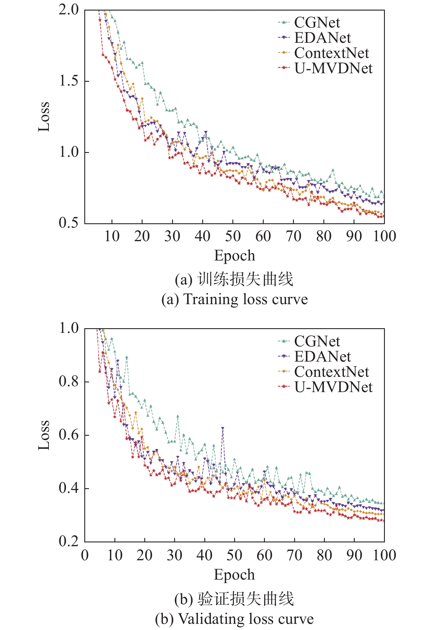
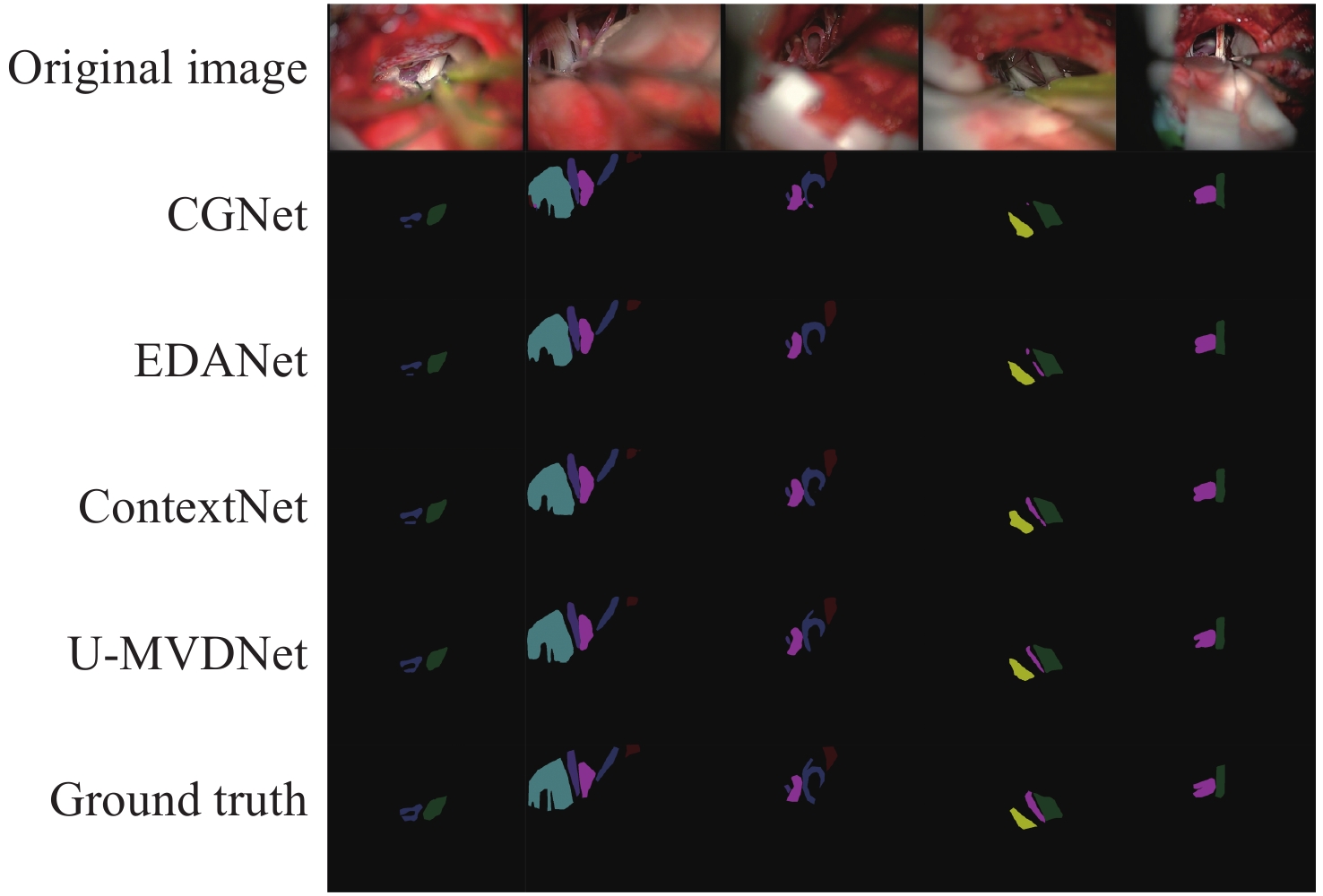
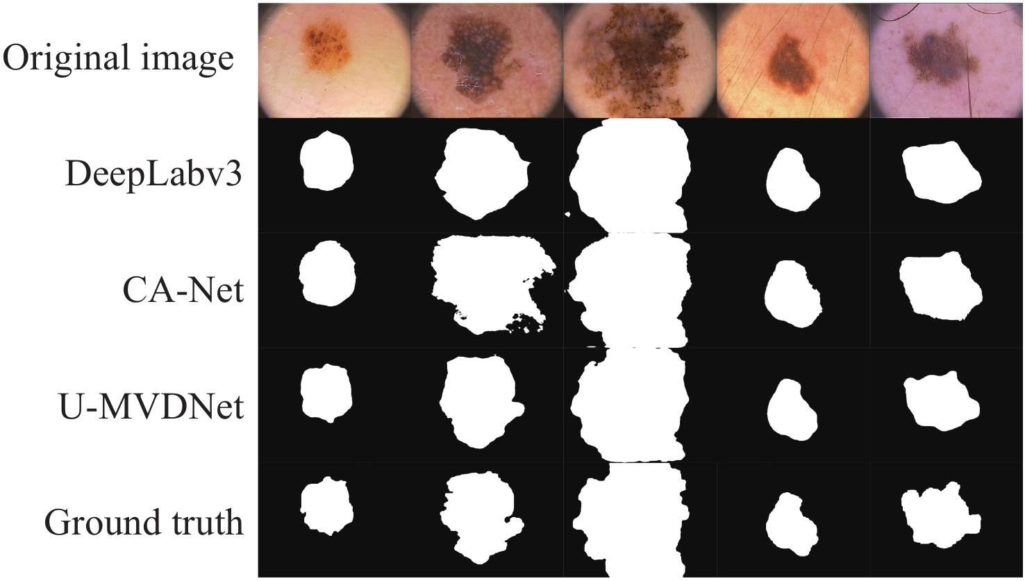
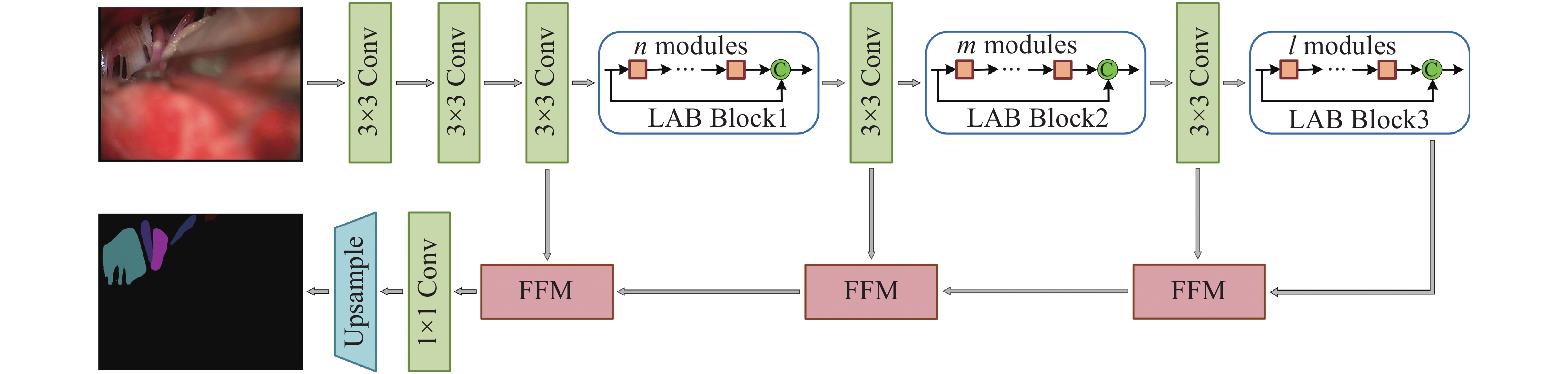
Aiming at the problems of large parameters and low semantic segmentation accuracy of real-time semantic segmentation networks for true-color microvascular decompression (MVD) images. This paper proposes a U-shaped lightweight fast semantic segmentation network U-MVDNet (U-Shaped Microvascular Decompression Network) for MVD scenarios, which consists of encoder-decoder structure. A Light Asymmetric Bottleneck Module (LABM) is designed in the encoder to encode context features. Feature Fusion Module (FFM) is introduced in the decoder to effectively combine high-level semantic features and underlying spatial details. Experimental results show that for the MVD test set, U-MVDNet achieves 0.66 M parameters, 76.29% mIoU (mean Intersection-over-Union), and 140 frame/s speed on NVIDIA GTX 2080Ti. And when input image size is 640 × 480, the real-time (24 frame/s) semantic segmentation is realized on NVIDIA Jetson AGX Xavier embedded development board. The proposed network has no pretrained model, fewer parameters, and fast inference speed. The semantic segmentation performance is superior to other comparison methods, and a good trade-off between segmentation accuracy and speed is achieved. Furthermore, U-MVDNet can also be easily developed and applied on embedded platform with superior performance and easy deployment.

| [1] |
BENNETTO L, PATEL N K, FULLER G. Trigeminal neuralgia and its management[J]. BMJ, 2007, 334(7586): 201-205. doi: 10.1136/bmj.39085.614792.BE
|
| [2] |
KIZILTAN M E, GUNDUZ A. Reorganization of sensory input at brainstem in hemifacial spasm and postparalytic facial syndrome[J]. Neurological Sciences, 2018, 39(2): 313-319. doi: 10.1007/s10072-017-3185-1
|
| [3] |
NAZIR A, CHEEMA M N, SHENG B, et al. OFF-eNET: an optimally fused fully end-to-end network for automatic dense volumetric 3D intracranial blood vessels segmentation[J]. IEEE Transactions on Image Processing, 2020, 29: 7192-7202. doi: 10.1109/TIP.2020.2999854
|
| [4] |
吴红宇, 郑波. 脑动静脉畸形CTA、DSA的影像学表现及诊断的对照性研究[J]. 中国CT和MRI杂志,2021,19(1):36-37,52. doi: 10.3969/j.issn.1672-5131.2021.01.012
WU H Y, ZHENG B. Analysis on imaging manifestations and diagnostic contrast of cerebral arteriovenous malformations in CTA and DSA[J]. Chinese Journal of CT and MRI, 2021, 19(1): 36-37,52. (in Chinese) doi: 10.3969/j.issn.1672-5131.2021.01.012
|
| [5] |
PATEL T R, PALIWAL N, JAISWAL P, et al. Multi-resolution CNN for brain vessel segmentation from cerebrovascular images of intracranial aneurysm: a comparison of U-Net and DeepMedic[J]. Proceedings of SPIE, 2020, 11314: 113142W.
|
| [6] |
王华. 磁共振血管成像与三维动脉自旋标记脑灌注成像技术诊断缺血性脑血管疾病一致性比较[J]. 实用医院临床杂志,2020,17(1):36-39. doi: 10.3969/j.issn.1672-6170.2020.01.011
WANG H. Comparison of consistency of MRA and 3D-ASL cerebral perfusion imaging in the diagnosisof ischemic cerebrovascular diseases[J]. Practical Journal of Clinical Medicine, 2020, 17(1): 36-39. (in Chinese) doi: 10.3969/j.issn.1672-6170.2020.01.011
|
| [7] |
徐冰洁. 电子计算机断层扫描联合核磁共振血管成像对脑血管疾病的诊断价值[J]. 临床合理用药杂志,2021,14(32):177-178. doi: 10.15887/j.cnki.13-1389/r.2021.32.071
XU B J. Diagnostic value of computed tomography combined with nuclear magnetic resonance angiography in cerebrovascular diseases[J]. Chinese Journal of Clinical Rational Drug Use, 2021, 14(32): 177-178. (in Chinese) doi: 10.15887/j.cnki.13-1389/r.2021.32.071
|
| [8] |
ZHANG H, XIA L K, SONG R, et al. . Cerebrovascular segmentation in mra via reverse edge attention network[C]. Proceedings of the 23rd International Conference on Medical Image Computing and Computer-Assisted Intervention, Springer, 2020: 66-75.
|
| [9] |
GUO X Y, XIAO R X, LU Y Y, et al. Cerebrovascular segmentation from TOF-MRA based on multiple-U-net with focal loss function[J]. Computer Methods and Programs in Biomedicine, 2021, 202: 105998. doi: 10.1016/j.cmpb.2021.105998
|
| [10] |
HILBERT A, MADAI V I, AKAY E M, et al. BRAVE-NET: fully automated arterial brain vessel segmentation in patients with cerebrovascular disease[J]. Frontiers in Artificial Intelligence, 2020, 3: 552258. doi: 10.3389/frai.2020.552258
|
| [11] |
陈星, 宋智洋, 周明全, 等. 面向脑血管分割的改进型非局部均值滤波算法研究[J]. 中国光学,2014,7(4):572-580.
CHEN X, SONG ZH Y, ZHOU M Q, et al. An improved non-local mean filter algorithm facing the cerebrovascular segmentation[J]. Chinese Optics, 2014, 7(4): 572-580. (in Chinese)
|
| [12] |
WANG R, LI CH, WANG J, et al. Threshold segmentation algorithm for automatic extraction of cerebral vessels from brain magnetic resonance angiography images[J]. Journal of Neuroscience Methods, 2015, 241: 30-36. doi: 10.1016/j.jneumeth.2014.12.003
|
| [13] |
BHUIYAN A, NATH B, CHUA J. An adaptive region growing segmentation for blood vessel detection from retinal images[C]. Visapp: Second International Conference on Computer Vision Theory and Applications, 2007: 404-409.
|
| [14] |
王醒策, 张美霞, 武仲科, 等. 基于全局LBF水平集模型的脑血管层次粗分割[J]. 光学 精密工程,2013,21(12):3283-3297. doi: 10.3788/OPE.20132112.3283
WANG X C, ZHANG M X, WU ZH K, et al. Level coarse brain vessel segmentation based on global LBF model[J]. Optics and Precision Engineering, 2013, 21(12): 3283-3297. (in Chinese) doi: 10.3788/OPE.20132112.3283
|
| [15] |
WANG J X, ZHAO SH F, LIU Z F, et al. An active contour model based on adaptive threshold for extraction of cerebral vascular structures[J]. Computational and Mathematical Methods in Medicine, 2016, 2016: 6472397.
|
| [16] |
陈晓冬, 艾大航, 张佳琛, 等. Gabor滤波融合卷积神经网络的路面裂缝检测方法[J]. 中国光学,2020,13(6):1293-1301. doi: 10.37188/CO.2020-0041
CHEN X D, AI D H, ZHANG J CH, et al. Gabor filter fusion network for pavement crack detection[J]. Chinese Optics, 2020, 13(6): 1293-1301. (in Chinese) doi: 10.37188/CO.2020-0041
|
| [17] |
王春哲, 安军社, 姜秀杰, 等. 基于卷积神经网络的候选区域优化算法[J]. 中国光学,2019,12(6):1348-1361. doi: 10.3788/co.20191206.1348
WANG CH ZH, AN J SH, JIANG X J, et al. Region proposal optimization algorithm based on convolutional neural networks[J]. Chinese Optics, 2019, 12(6): 1348-1361. (in Chinese) doi: 10.3788/co.20191206.1348
|
| [18] |
LONG J, SHELHAMER E, DARRELL T. Fully convolutional networks for semantic segmentation[C]. Proceedings of 2015 IEEE Conference on Computer Vision and Pattern Recognition, IEEE, 2015: 3431-3440.
|
| [19] |
SHVETS A A, IGLOVIKOV V I, RAKHLIN A, et al. . Angiodysplasia detection and localization using deep convolutional neural networks[C]. Proceedings of 2018 17th IEEE International Conference on Machine Learning and Applications (ICMLA), IEEE, 2018: 612-617.
|
| [20] |
ZHANG M, ZHANG CH, WU X, et al. A neural network approach to segment brain blood vessels in digital subtraction angiography[J]. Computer Methods Programs in Biomedicine, 2020, 185: 105159. doi: 10.1016/j.cmpb.2019.105159
|
| [21] |
XIA L K, XIE Y X, WANG Q W, et al. A nested parallel multiscale convolution for cerebrovascular segmentation[J]. Medical Physics, 2021, 48(12): 7971-7983. doi: 10.1002/mp.15280
|
| [22] |
MENG C, SUN K, GUAN SH Y, et al. Multiscale dense convolutional neural network for DSA cerebrovascular segmentation[J]. Neurocomputing, 2020, 373: 123-134. doi: 10.1016/j.neucom.2019.10.035
|
| [23] |
HUANG G, LIU ZH, VAN DER MAATEN L, et al. . Densely connected convolutional networks[C]. Proceedings of 2017 IEEE Conference on Computer Vision and Pattern Recognition, IEEE, 2017: 2261-2269.
|
| [24] |
ALVAREZ J, PETERSSON L. DecomposeMe: simplifying convnets for end-to-end learning[J]. arXiv preprint arXiv:
|
| [25] |
HE K M, ZHANG X Y, REN SH Q, et al. . Delving deep into rectifiers: Surpassing human-level performance on imagenet classification[C]. Proceedings of 2015 IEEE International Conference on Computer Vision, IEEE, 2015: 1026-1034.
|
| [26] |
CODELLA N C F, GUTMAN D, CELEBI M E, et al. . Skin lesion analysis toward melanoma detection: a challenge at the 2017 international symposium on biomedical imaging (ISBI), hosted by the international skin imaging collaboration (ISIC)[C]. Proceedings of the 15th International Symposium on Biomedical Imaging, IEEE, 2016.
|
| [27] |
MENDONÇA T, FERREIRA P M, MARQUES J S, et al. . PH2-A dermoscopic image database for research and benchmarking[C]. Proceedings of the 35th Annual International Conference of the IEEE Engineering in Medicine and Biology Society (EMBC), IEEE, 2013: 5437-5440.
|
| [28] |
WU T Y, TANG SH, ZHANG R, et al. CGNet: a light-weight context guided network for semantic segmentation[J]. IEEE Transactions on Image Processing, 2020, 30: 1169-1179.
|
| [29] |
LO S Y, HANG H M, CHAN SH W, et al. . Efficient dense modules of asymmetric convolution for real-time semantic segmentation[C]. Proceedings of the ACM Multimedia Asia, ACM, 2019: 1.
|
| [30] |
POUDEL R P K, BONDE U, LIWICKI S, et al. ContextNet: exploring context and detail for semantic segmentation in real-time[J]. arXiv preprint arXiv:
|
| [31] |
CHEN L C, PAPANDREOU G, SCHROFF F, et al. Rethinking atrous convolution for semantic image segmentation[J]. arXiv preprint arXiv:
|
| [32] |
GU R, WANG G T, SONG T, et al. CA-Net: comprehensive attention convolutional neural networks for explainable medical image segmentation[J]. IEEE Transactions on Medical Imaging, 2021, 40(2): 699-711. doi: 10.1109/TMI.2020.3035253
|
Figures(6) / Tables(12)


 DownLoad: