| Citation: | WANG Peng, ZHOU Yao, ZHAO Yu-xuan, FEI Peng. Double-ring-modulated light sheet fluorescence microscopic technique for multi-scale high-resolution 3D imaging[J]. Chinese Optics, 2022, 15(6): 1321-1331. doi: 10.37188/CO.2022-0093

|
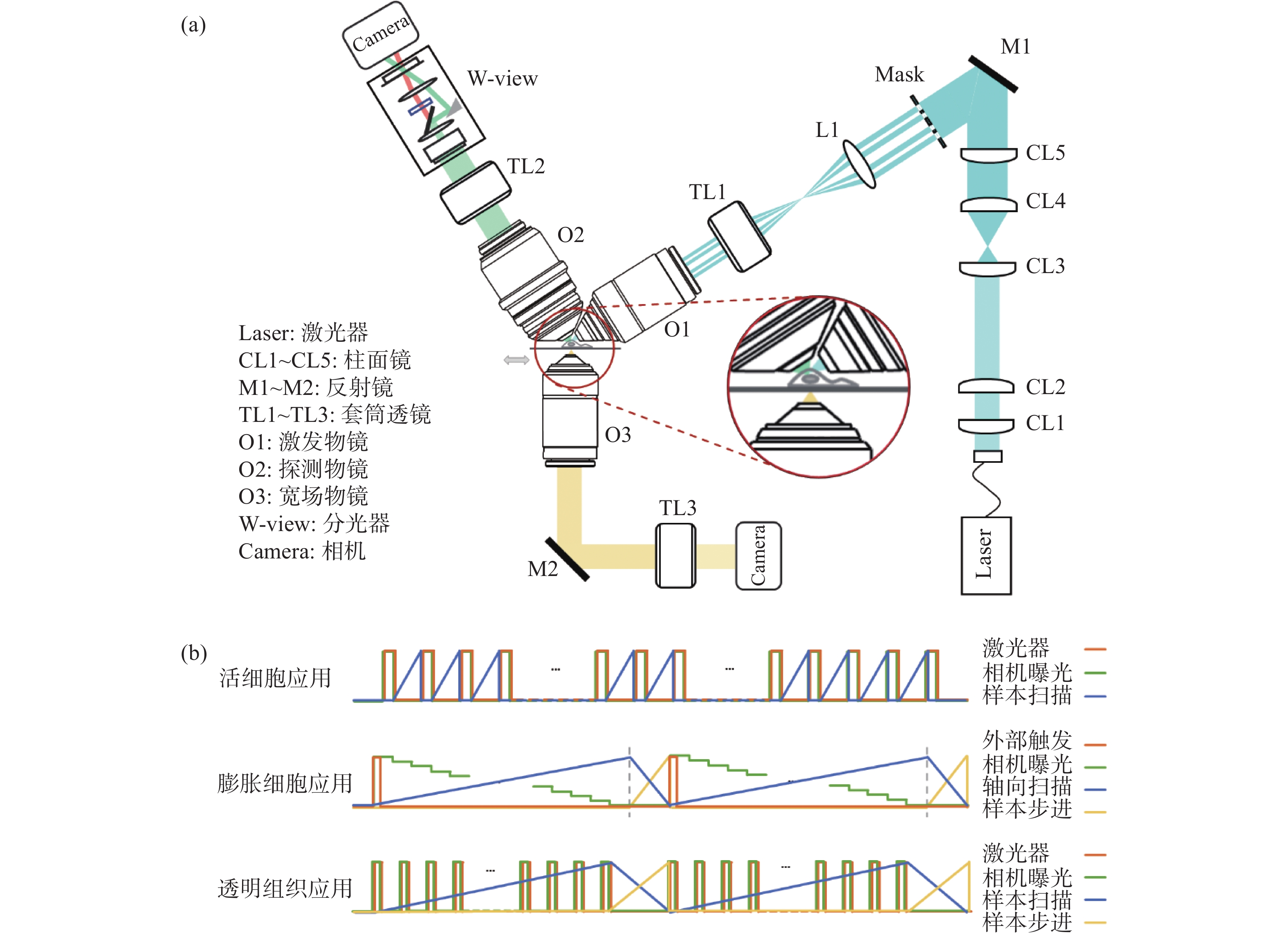
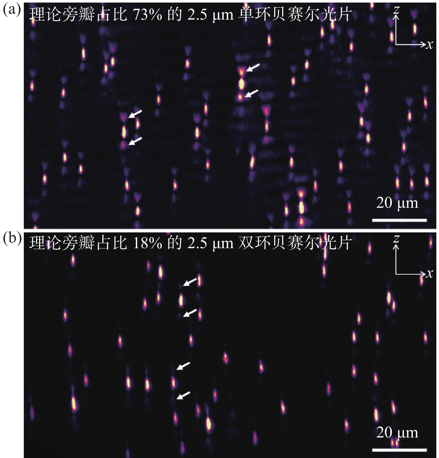
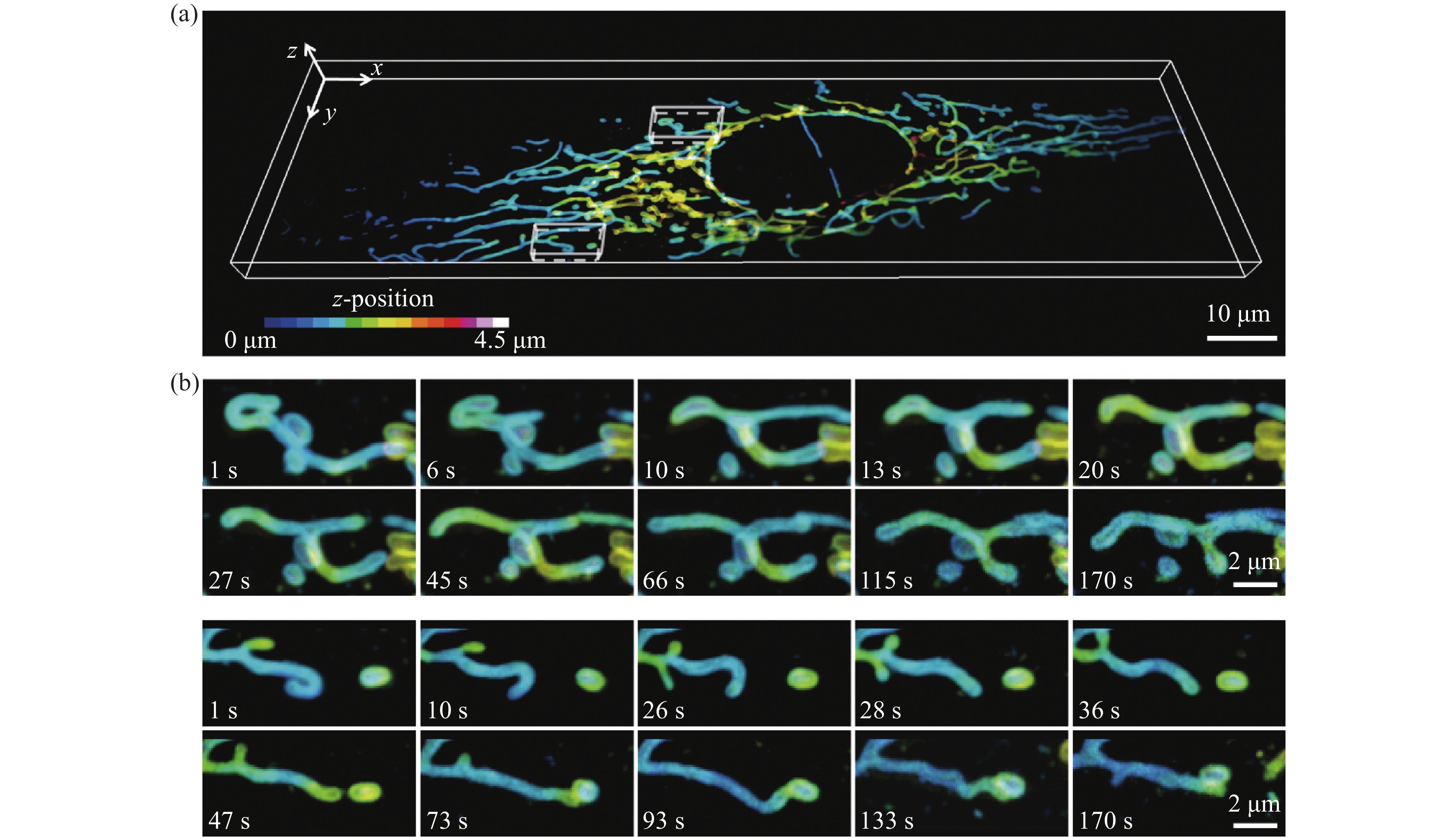
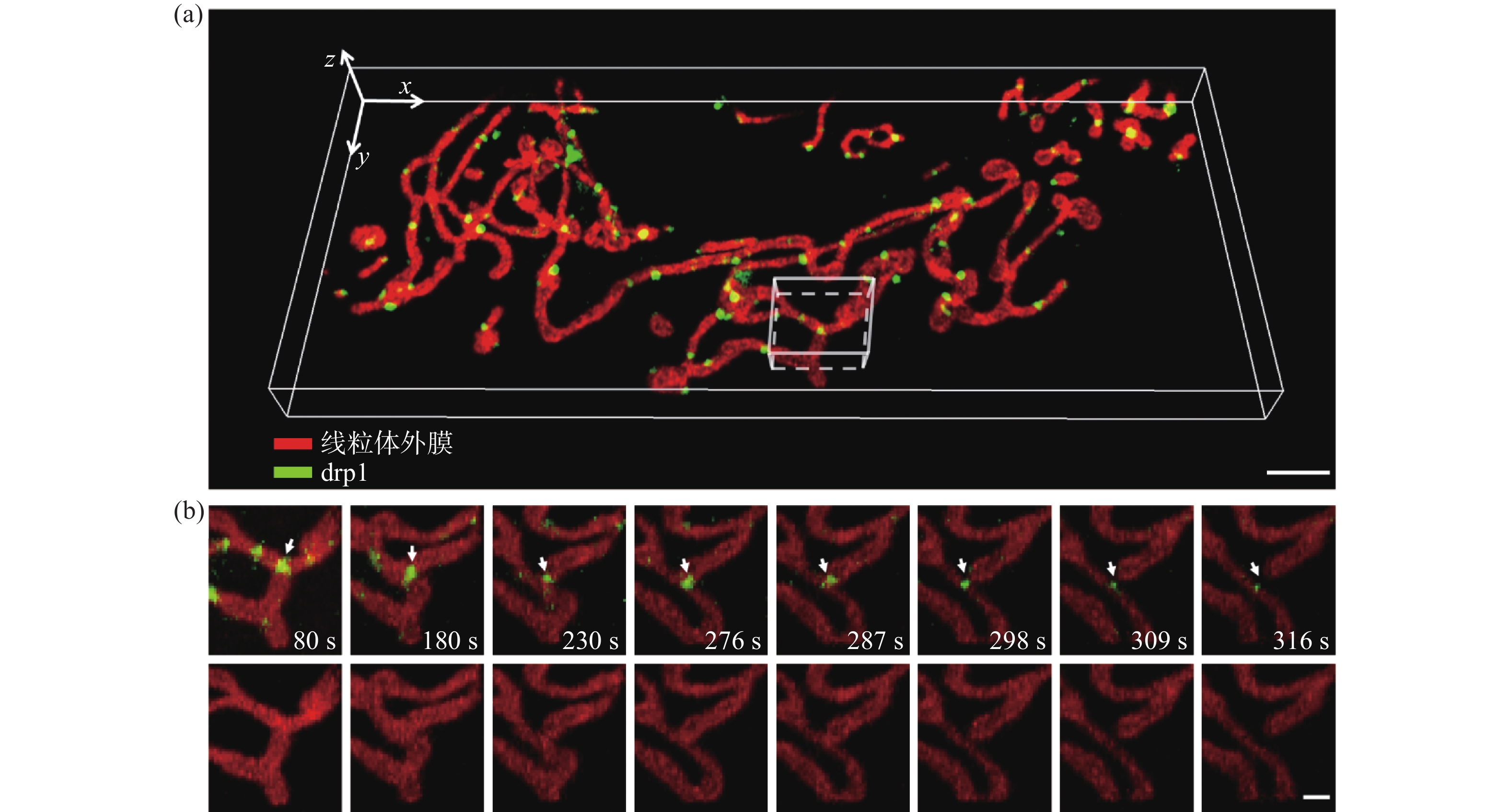
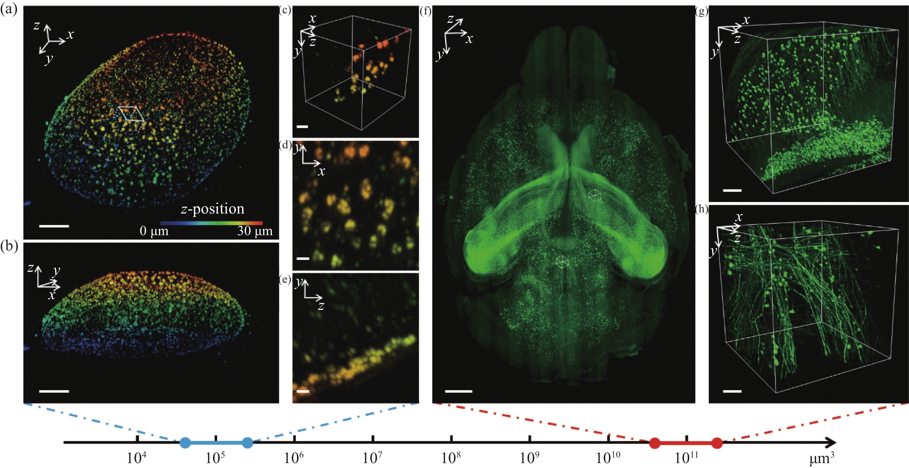
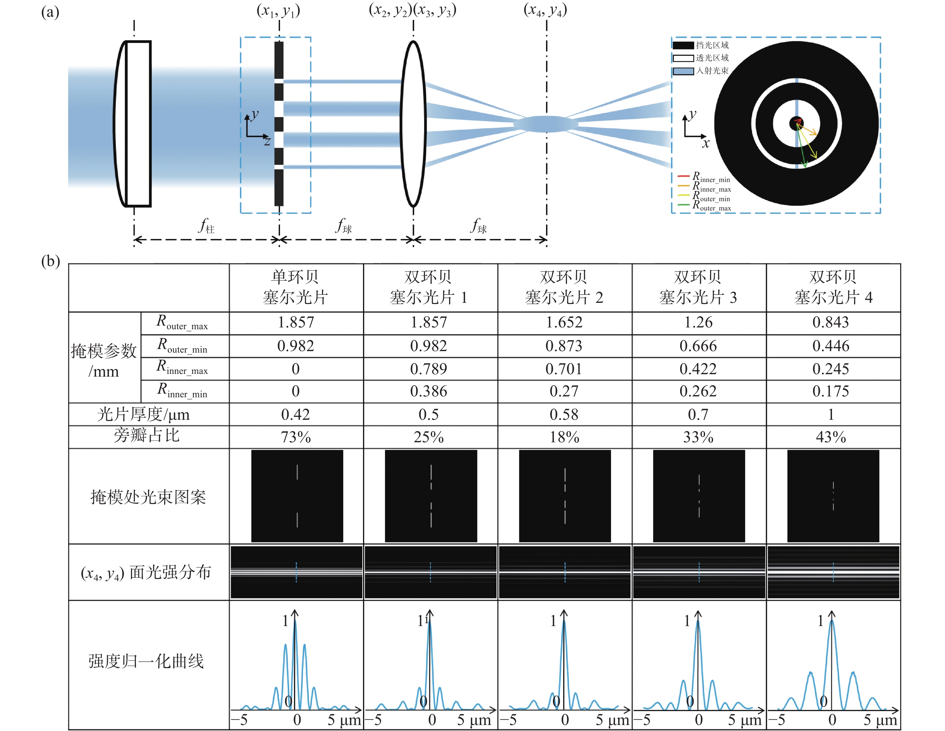
In this paper, we propose a non-diffraction Light Sheet Fluorescence Microscopy (LSFM) technique, which readily enables multi-scale 3D fluorescence imaging of diverse biological samples with size ranging from microns to centimeters. To solve the problem of heavy sidelobes in conventional non-diffraction Bessel LSFM, we invent a double-ring-modulated approach which can generate non-diffraction light sheets with ~0.4 to ~5 µm tunable thickness and the ratio of the sidelobe was reduced to less than 30%. Then we built a multi-scale LSFM system based on this novel approach. The system showed versatile multi-scale imaging abilities, such as dual-color 3D dynamic imaging of single live cell, 3D super-resolution imaging of expansion cells and high-throughput 3D mapping of entire meso-scale organs. Therefore, we demonstrate that this multi-scale imaging modality can substantially improve the efficiency of LSFM for advancing various biomedical studies, such as cell biology, tissue pathology, and neuroscience.

| [1] |
LICHTMAN J W, CONCHELLO J A. Fluorescence microscopy[J]. Nature Methods, 2005, 2(12): 910-919. doi: 10.1038/nmeth817
|
| [2] |
CHOQUET D, SAINLOS M, SIBARITA J B. Advanced imaging and labelling methods to decipher brain cell organization and function[J]. Nature Reviews Neuroscience, 2021, 22(4): 237-255. doi: 10.1038/s41583-021-00441-z
|
| [3] |
STELZER E H K, STROBL F, CHANG B J, et al. Light sheet fluorescence microscopy[J]. Nature Reviews Methods Primers, 2021, 1(1): 73. doi: 10.1038/s43586-021-00069-4
|
| [4] |
HUISKEN J, SWOGER J, DEL BENE F, et al. Optical sectioning deep inside live embryos by selective plane illumination microscopy[J]. Science, 2004, 305(5686): 1007-1009. doi: 10.1126/science.1100035
|
| [5] |
KELLER P J, SCHMIDT A D, WITTBRODT J, et al. Reconstruction of zebrafish early embryonic development by scanned light sheet microscopy[J]. Science, 2008, 322(5904): 1065-1069. doi: 10.1126/science.1162493
|
| [6] |
DEAN K M, ROUDOT P, WELF E S, et al. Deconvolution-free subcellular imaging with axially swept light sheet microscopy[J]. Biophysical Journal, 2015, 108(12): 2807-2815. doi: 10.1016/j.bpj.2015.05.013
|
| [7] |
FAHRBACH F O, GURCHENKOV V, ALESSANDRI K, et al. Self-reconstructing sectioned Bessel beams offer submicron optical sectioning for large fields of view in light-sheet microscopy[J]. Optics Express, 2013, 21(9): 11425-11440. doi: 10.1364/OE.21.011425
|
| [8] |
LIU Y CH, KE Y G, ZHOU J X, et al. Generation of perfect vortex and vector beams based on Pancharatnam-Berry phase elements[J]. Scientific Reports, 2017, 7(1): 44096. doi: 10.1038/srep44096
|
| [9] |
ZHAO T, LAU S C, WANG Y, et al. Multicolor 4D fluorescence microscopy using ultrathin bessel light sheets[J]. Scientific Reports, 2016, 6(1): 26159. doi: 10.1038/srep26159
|
| [10] |
CAO B, COELHO S, LI J R, et al. Volumetric interferometric lattice light-sheet imaging[J]. Nature Biotechnology, 2021, 39(11): 1385-1393. doi: 10.1038/s41587-021-01042-y
|
| [11] |
CHANG B J, KITTISOPIKUL M, DEAN K M, et al. Universal light-sheet generation with field synthesis[J]. Nature Methods, 2019, 16(3): 235-238. doi: 10.1038/s41592-019-0327-9
|
| [12] |
DUNSBY C. Optically sectioned imaging by oblique plane microscopy[J]. Optics Express, 2008, 16(25): 20306-20316. doi: 10.1364/OE.16.020306
|
| [13] |
YANG B, LANGE M, MILLETT-SIKKING A, et al. DaXi—high-resolution, large imaging volume and multi-view single-objective light-sheet microscopy[J]. Nature Methods, 2022, 19(4): 461-469. doi: 10.1038/s41592-022-01417-2
|
| [14] |
POWER R M, HUISKEN J. A guide to light-sheet fluorescence microscopy for multiscale imaging[J]. Nature Methods, 2017, 14(4): 360-373. doi: 10.1038/nmeth.4224
|
| [15] |
MORI S. Side lobe suppression of a Bessel beam for high aspect ratio laser processing[J]. Precision Engineering, 2015, 39: 79-85. doi: 10.1016/j.precisioneng.2014.07.008
|
| [16] |
郁道银, 谈恒英. 工程光学[M]. 北京: 机械工业出版社, 2015.
YU D Y, TAN H Y. Engineering Optics[M]. Beijing: China Machine Press, 2015. (in Chinese)
|
| [17] |
ZHAO Y X, ZHANG M, ZHANG W T, et al. Isotropic super-resolution light-sheet microscopy of dynamic intracellular structures at subsecond timescales[J]. Nature Methods, 2022, 19(3): 359-369. doi: 10.1038/s41592-022-01395-5
|
| [18] |
SCHERMELLEH L, FERRAND A, HUSER T, et al. Super-resolution microscopy demystified[J]. Nature Cell Biology, 2019, 21(1): 72-84. doi: 10.1038/s41556-018-0251-8
|
| [19] |
WU Y C, HAN X F, SU Y J, et al. Multiview confocal super-resolution microscopy[J]. Nature, 2021, 600(7888): 279-284. doi: 10.1038/s41586-021-04110-0
|
| [20] |
VALLI J, GARCIA-BURGOS A, ROONEY L M, et al. Seeing beyond the limit: a guide to choosing the right super-resolution microscopy technique[J]. Journal of Biological Chemistry, 2021, 297(1): 100791. doi: 10.1016/j.jbc.2021.100791
|
| [21] |
ZHAO W S, ZHAO SH Q, LI L J, et al. Sparse deconvolution improves the resolution of live-cell super-resolution fluorescence microscopy[J]. Nature Biotechnology, 2022, 40(4): 606-617. doi: 10.1038/s41587-021-01092-2
|
| [22] |
HUANG X SH, FAN J CH, LI L J, et al. Fast, long-term, super-resolution imaging with Hessian structured illumination microscopy[J]. Nature Biotechnology, 2018, 36(5): 451-459. doi: 10.1038/nbt.4115
|
| [23] |
QIAO CH, LI D, GUO Y T, et al. Evaluation and development of deep neural networks for image super-resolution in optical microscopy[J]. Nature Methods, 2021, 18(2): 194-202. doi: 10.1038/s41592-020-01048-5
|
| [24] |
ZHELUDEV N I, YUAN G H. Optical superoscillation technologies beyond the diffraction limit[J]. Nature Reviews Physics, 2022, 4(1): 16-32. doi: 10.1038/s42254-021-00382-7
|
| [25] |
BODÉN A, PENNACCHIETTI F, COCEANO G, et al. Volumetric live cell imaging with three-dimensional parallelized RESOLFT microscopy[J]. Nature Biotechnology, 2021, 39(5): 609-618. doi: 10.1038/s41587-020-00779-2
|
| [26] |
SUN D E, FAN X Q, SHI Y J, et al. Click-ExM enables expansion microscopy for all biomolecules[J]. Nature Methods, 2021, 18(1): 107-113. doi: 10.1038/s41592-020-01005-2
|
| [27] |
GAO R X, YU C C, GAO L Y, et al. A highly homogeneous polymer composed of tetrahedron-like monomers for high-isotropy expansion microscopy[J]. Nature Nanotechnology, 2021, 16(6): 698-707. doi: 10.1038/s41565-021-00875-7
|
| [28] |
THEVATHASAN J V, KAHNWALD M, CIEŚLIŃSKI K, et al. Nuclear pores as versatile reference standards for quantitative superresolution microscopy[J]. Nature Methods, 2019, 16(10): 1045-1053. doi: 10.1038/s41592-019-0574-9
|
| [29] |
JING D, ZHANG SH W, LUO W J, et al. Tissue clearing of both hard and soft tissue organs with the pegasos method[J]. Cell Research, 2018, 28(8): 803-818. doi: 10.1038/s41422-018-0049-z
|
| [30] |
ZHU J T, LIU X M, DENG Y T, et al. Tissue optical clearing for 3D visualization of vascular networks: a review[J]. Vascular Pharmacology, 2021, 141: 106905. doi: 10.1016/j.vph.2021.106905
|
| [31] |
SUN Q T, LI X N, REN M, et al. A whole-brain map of long-range inputs to GABAergic interneurons in the mouse medial prefrontal cortex[J]. Nature Neuroscience, 2019, 22(8): 1357-1370. doi: 10.1038/s41593-019-0429-9
|
| [32] |
FANG CH Y, YU T T, CHU T T, et al. Minutes-timescale 3D isotropic imaging of entire organs at subcellular resolution by content-aware compressed-sensing light-sheet microscopy[J]. Nature Communications, 2021, 12(1): 107. doi: 10.1038/s41467-020-20329-3
|
| [33] |
XU F, SHEN Y, DING L F, et al. High-throughput mapping of a whole rhesus monkey brain at micrometer resolution[J]. Nature Biotechnology, 2021, 39(12): 1521-1528. doi: 10.1038/s41587-021-00986-5
|
Figures(6) / Tables(1)


 DownLoad: