| Citation: | LANG Song, ZHANG Yan-wei, ZHENG Han-qing, XU Lin-yu, WANG Lu-han, GONG Yan. Wide-field-of-view and high-resolution HiLo optical sectioning microscopy system[J]. Chinese Optics, 2022, 15(6): 1302-1312. doi: 10.37188/CO.2022-0087

|
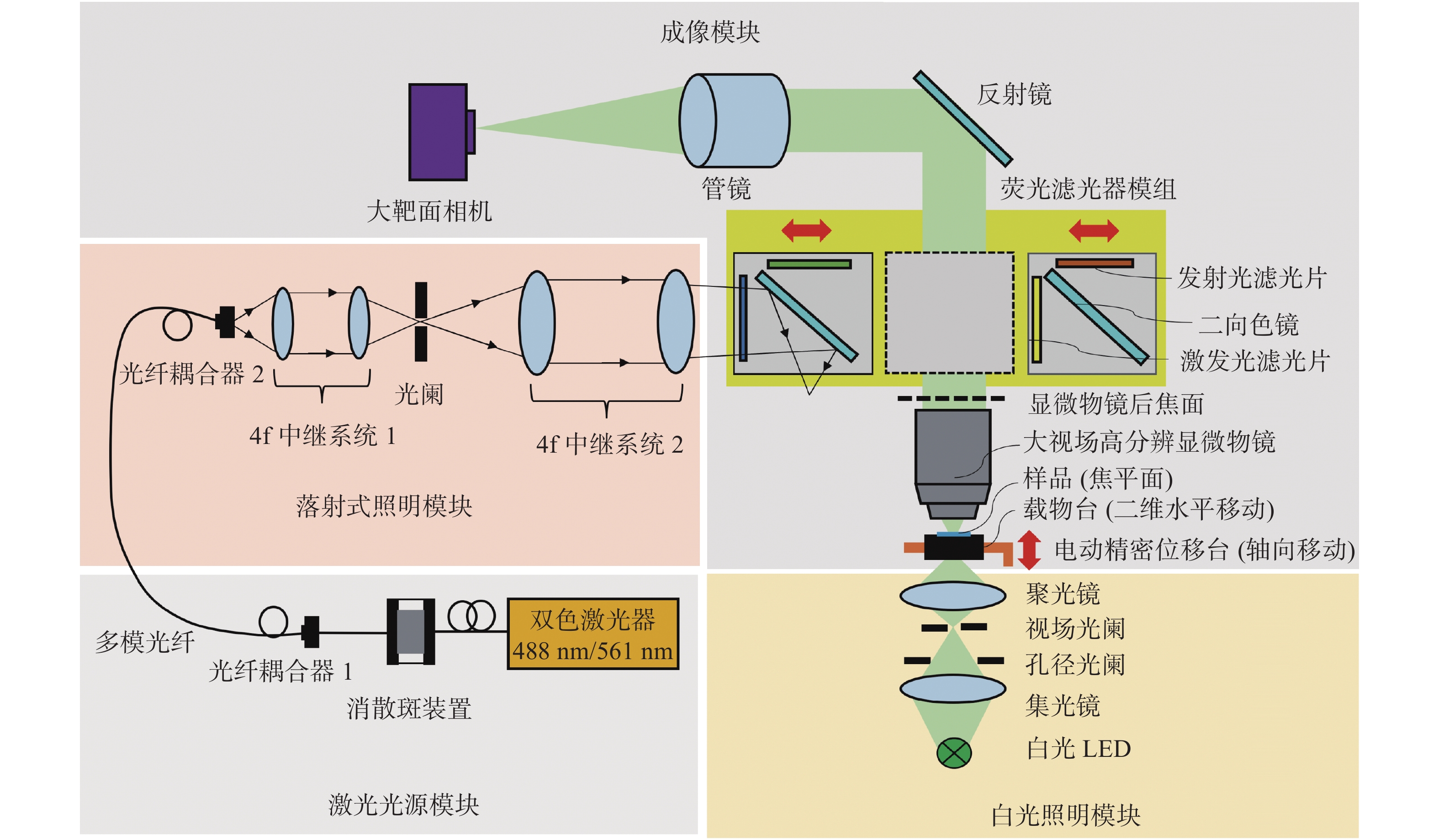
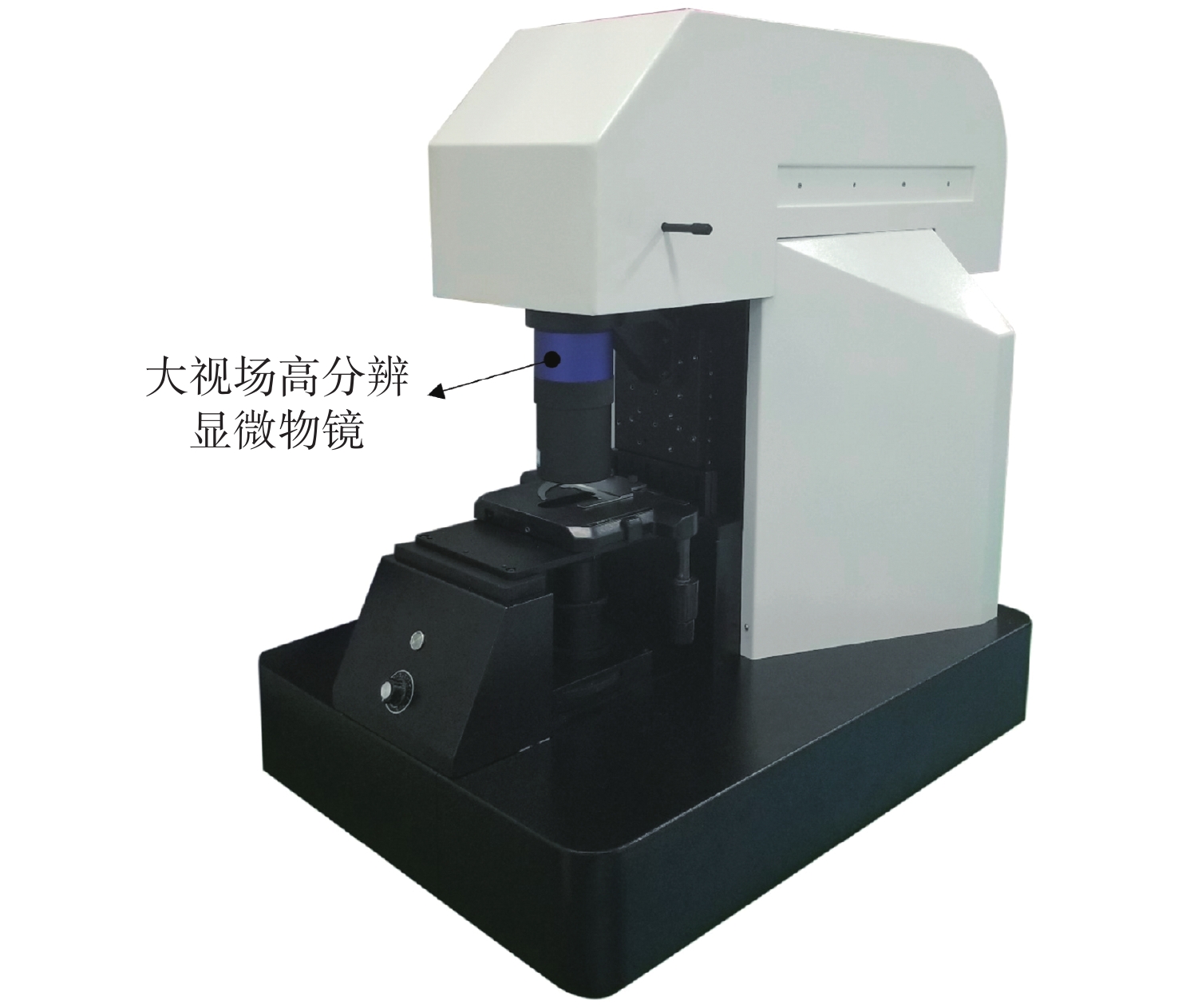
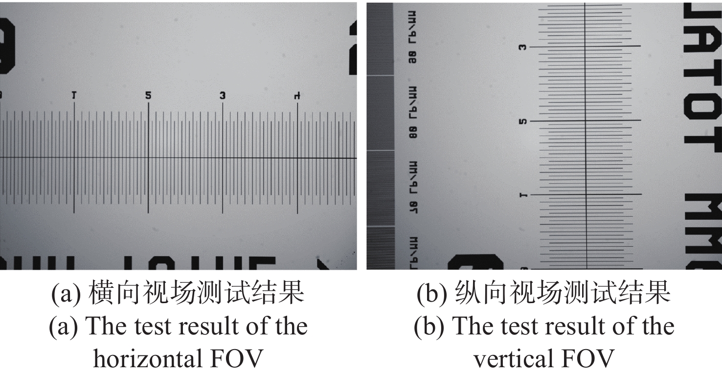
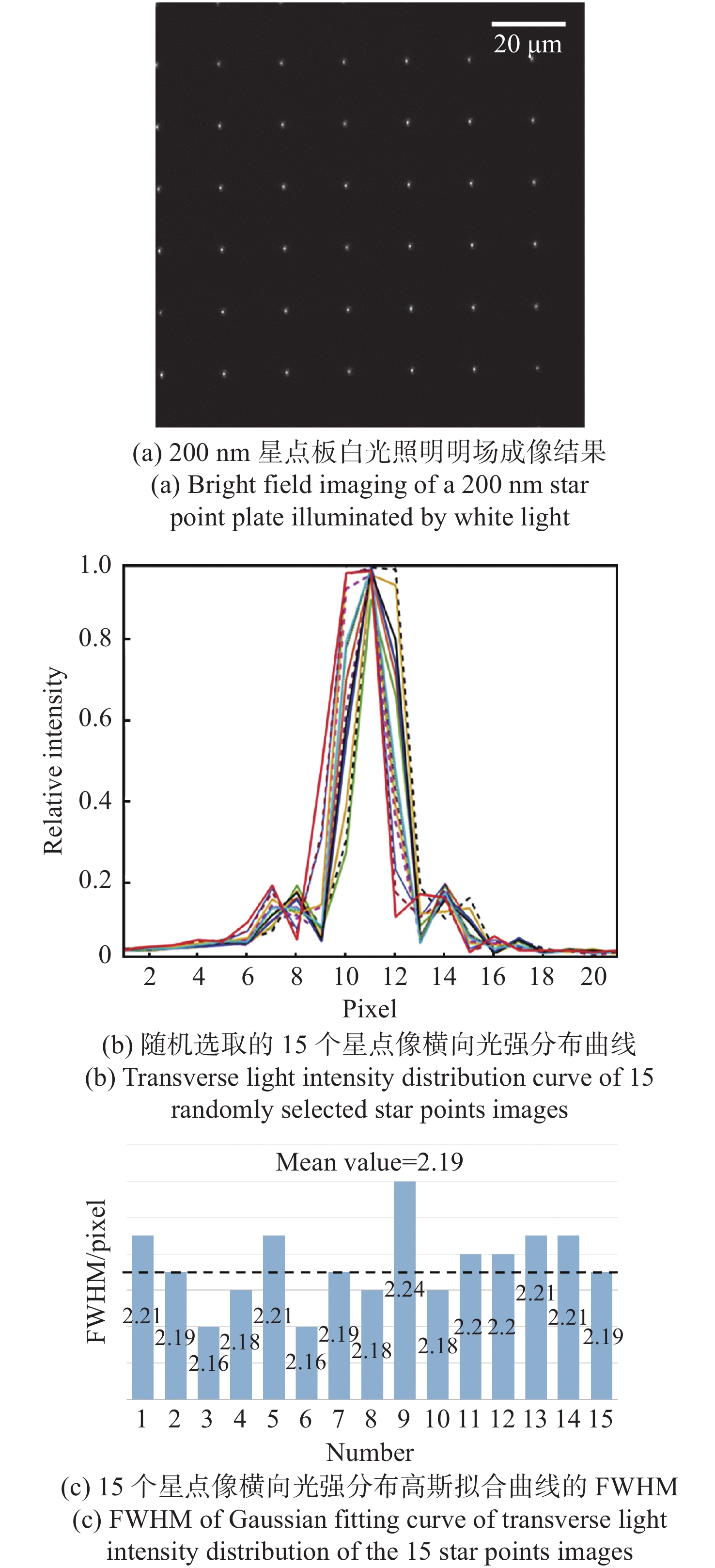
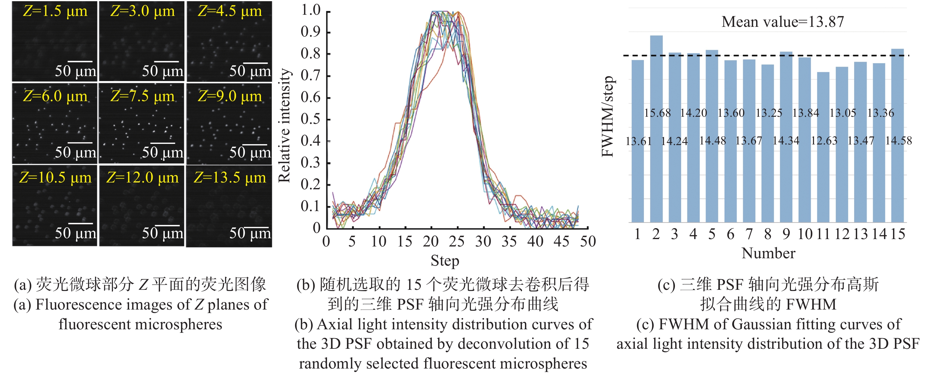
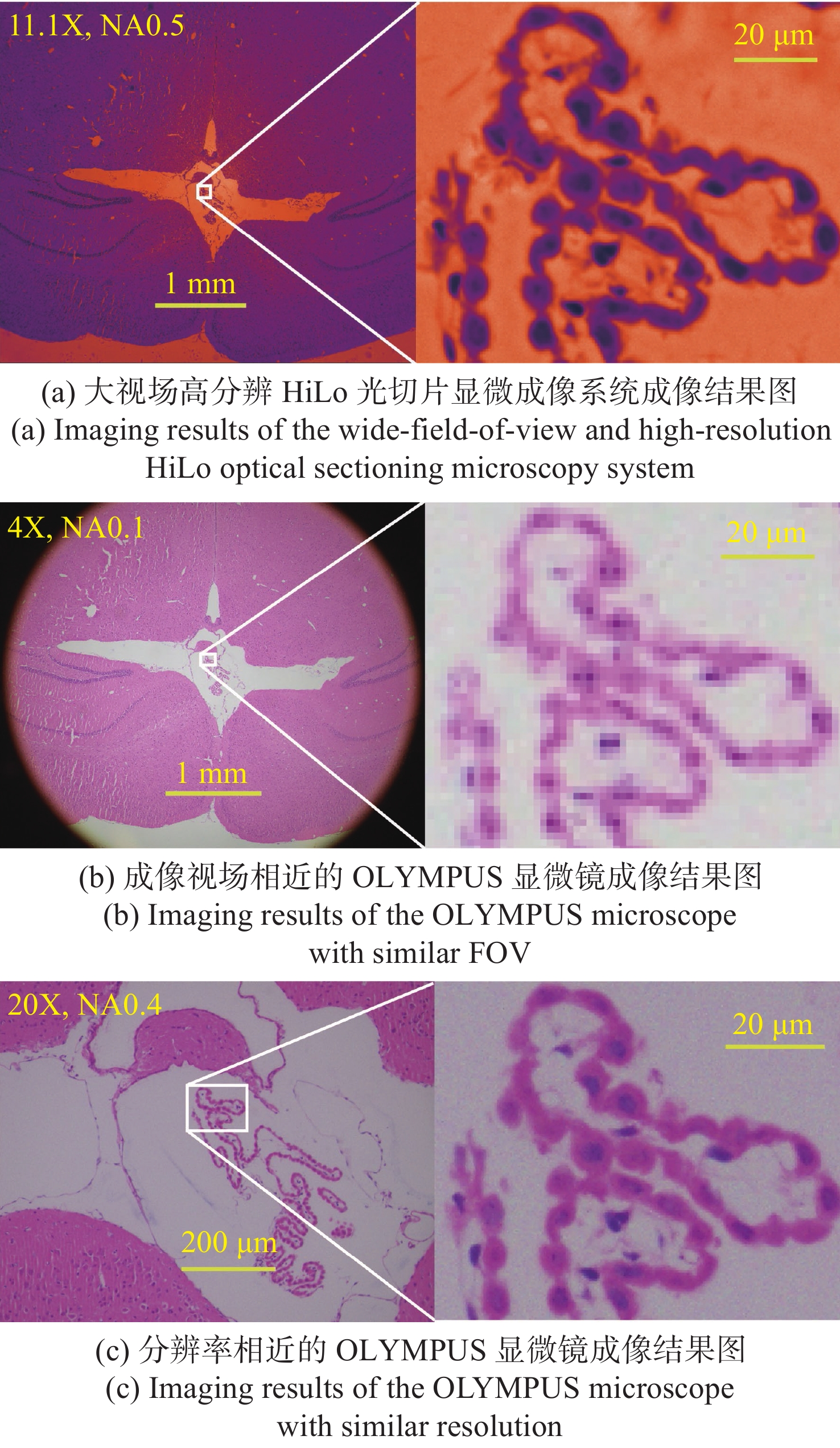
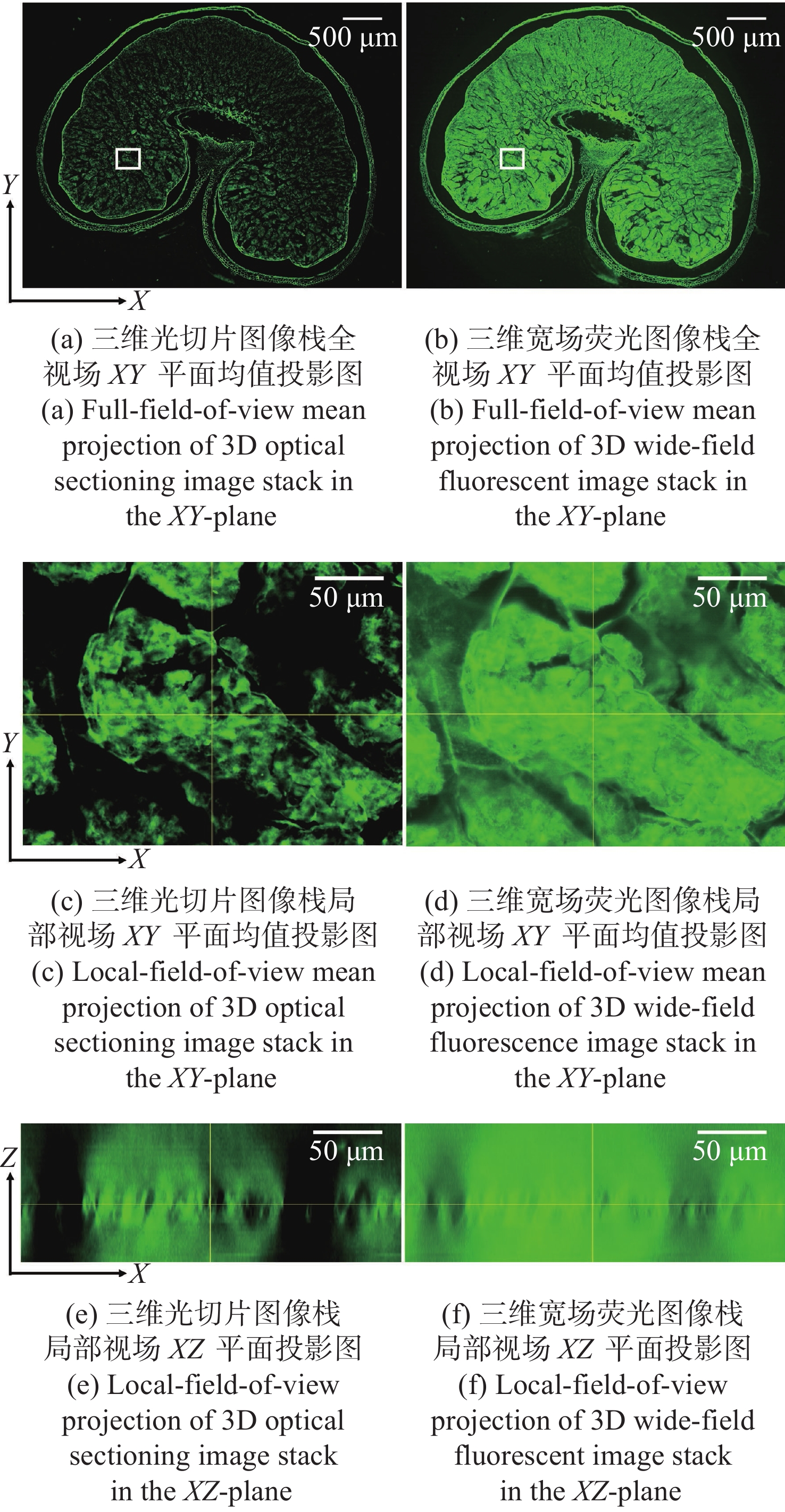
The fields of modern biology and biomedicine urgently need wide-field-of-view (FOV), high-resolution microscopic technology and instruments for trans-scale observation of biological samples to meet the requirement of major scientific for research. Limited by the spatial bandwidth product, traditional commercial microscopes cannot meet this demand. Besides, the existing high spatial bandwidth product microscopy systems have problems such as bulky volume and high implementation costs. In this paper, based on the HiLo optical sectioning technology and the self-designed wide-field-of-view and high-resolution objective, a wide-field-of-view and high-resolution HiLo optical sectioning microscopy system was developed. The FOV and imaging resolution of this system were tested. Brightfield imaging experiments were carried out on mouse brain slices by this system and the results were compared with that of OLYMPUS commercial microscope. At the same time, wide-field fluorescence imaging comparison experiments were carried out on wheat seed fluorescent slices. The experiment results show that the FOV of this system reaches 4.8 mm×3.6 mm (the diagonal FOV is 6.0 mm), the lateral resolution reaches 0.74 μm, and the axial resolution reaches 4.16 μm. The comparative experiment proved that this system has the advantages of wide FOV, high resolution and the ability of fast optical sectioning imaging simultaneously. This system can carry out rapid 3D imaging of large-volume biological samples, which will provide strong technical support for researches such as embryonic development, brain imaging, and digital pathology diagnosis.

| [1] |
骆清铭. 脑空间信息学——连接脑科学与类脑人工智能的桥梁[J]. 中国科学:生命科学,2017,47(10):1015-1024. doi: 10.1360/N052017-00094
LUO Q M. Brainsmatics—bridging the brain science and brain-inspired artificial intelligence[J]. Scientia Sinica Vitae, 2017, 47(10): 1015-1024. (in Chinese) doi: 10.1360/N052017-00094
|
| [2] |
QU L, LI Y, XIE P, et al. Cross-modal coherent registration of whole mouse brains[J]. Nature Methods, 2022, 19(1): 111-118. doi: 10.1038/s41592-021-01334-w
|
| [3] |
YU W, KANG L, TSANG V T C, et al.. Three-dimensional multicolor subcellular imaging by fast serial sectioning tomography for centimeter-scale specimens[J]. Biorxiv, 2021,doi: 10.1101/2021.11.11.468237.
|
| [4] |
BERTELS S, JAGGY M, RICHTER B, et al. Geometrically defined environments direct cell division rate and subcellular YAP localization in single mouse embryonic stem cells[J]. Scientific Reports, 2021, 11(1): 9269. doi: 10.1038/s41598-021-88336-y
|
| [5] |
WU J M, LU ZH, JIANG D, et al. Iterative tomography with digital adaptive optics permits hour-long intravital observation of 3D subcellular dynamics at millisecond scale[J]. Cell, 2021, 184(12): 3318-3332.e17. doi: 10.1016/j.cell.2021.04.029
|
| [6] |
HUGONNET H, KIM Y W, LEE M, et al. Multiscale label-free volumetric holographic histopathology of thick-tissue slides with subcellular resolution[J]. Advanced Photonics, 2021, 3(2): 026004.
|
| [7] |
LI A N, GONG H, ZHANG B, et al. Micro-optical sectioning tomography to obtain a high-resolution atlas of the mouse brain[J]. Science, 2010, 330(6009): 1404-1408. doi: 10.1126/science.1191776
|
| [8] |
ZHANG Y, KANG L, YU W T, et al. Three-dimensional label-free histological imaging of whole organs by microtomy-assisted autofluorescence tomography[J]. iScience, 2022, 25(1): 103721. doi: 10.1016/j.isci.2021.103721
|
| [9] |
TSAI P S, MATEO C, FIELD J J, et al. Ultra-large field-of-view two-photon microscopy[J]. Optics Express, 2015, 23(11): 13833-13847. doi: 10.1364/OE.23.013833
|
| [10] |
ZHONG Q Y, JIANG CH Y, ZHANG D J, et al. High-throughput optical sectioning via line-scanning imaging with digital structured modulation[J]. Optics Letters, 2021, 46(3): 504-507. doi: 10.1364/OL.412323
|
| [11] |
ZHENG G A, HORSTMEYER R, YANG CH H. Wide-field, high-resolution Fourier ptychographic microscopy[J]. Nature Photonics, 2013, 7(9): 739-745. doi: 10.1038/nphoton.2013.187
|
| [12] |
ZHENG G A, SHEN CH, JIANG SH W, et al. Concept, implementations and applications of Fourier ptychography[J]. Nature Reviews Physics, 2021, 3(3): 207-223. doi: 10.1038/s42254-021-00280-y
|
| [13] |
FAN J T, SUO J L, WU J M, et al. Video-rate imaging of biological dynamics at centimetre scale and micrometre resolution[J]. Nature Photonics, 2019, 13(11): 809-816. doi: 10.1038/s41566-019-0474-7
|
| [14] |
MCCONNELL G, TRÄGÅRDH J, AMOR R, et al. A novel optical microscope for imaging large embryos and tissue volumes with sub-cellular resolution throughout[J]. Elife, 2016, 5: e18659. doi: 10.7554/eLife.18659
|
| [15] |
MCCONNELL G, AMOS W B. Application of the mesolens for subcellular resolution imaging of intact larval and whole adult Drosophila[J]. Journal of Microscopy, 2018, 270(2): 252-258. doi: 10.1111/jmi.12693
|
| [16] |
SOFRONIEW N J, FLICKINGER D, KING J, et al. A large field of view two-photon mesoscope with subcellular resolution for in vivo imaging[J]. Elife, 2016, 5: e14472. doi: 10.7554/eLife.14472
|
| [17] |
YU CH H, STIRMAN J N, YU Y Y, et al. Diesel2p mesoscope with dual independent scan engines for flexible capture of dynamics in distributed neural circuitry[J]. Nature Communications, 2021, 12(1): 6639. doi: 10.1038/s41467-021-26736-4
|
| [18] |
张小宇. 基于深度学习的显微光学层析[D]. 武汉: 华中科技大学, 2020.
ZHANG X Y. Deep learning-based optical sectioning microscopy[D]. Wuhan: Huazhong University of Science and Technology, 2020. (in Chinese)
|
| [19] |
YANG M K, ZHOU ZH Q, ZHANG J X, et al. MATRIEX imaging: multiarea two-photon real-time in vivo explorer[J]. Light:Science &Applications, 2019, 8: 109.
|
| [20] |
STELZER E H K, STROBL F, CHANG B J, et al. Light sheet fluorescence microscopy[J]. Nature Reviews Methods Primers, 2021, 1(1): 73. doi: 10.1038/s43586-021-00069-4
|
| [21] |
PRIYADARSHI A, DULLO F T, WOLFSON D L, et al. A transparent waveguide chip for versatile total internal reflection fluorescence-based microscopy and nanoscopy[J]. Communications Materials, 2021, 2(1): 85. doi: 10.1038/s43246-021-00192-5
|
| [22] |
NWANESHIUDU A, KUSCHAL C, SAKAMOTO F H, et al. Introduction to confocal microscopy[J]. Journal of Investigative Dermatology, 2012, 132(12): 1-5. doi: 10.1038/jid.2012.429
|
| [23] |
XU L Y, ZHANG Y W, LANG S, et al. Structured illumination microscopy based on asymmetric three-beam interference[J]. Journal of Innovative Optical Health Sciences, 2021, 14(2): 2050027. doi: 10.1142/S1793545820500273
|
| [24] |
尹君, 王少飞, 张俊杰, 等. 基于动态散斑照明的宽场荧光显微技术理论研究[J]. 物理学报,2021,70(23):238701. doi: 10.7498/aps.70.20211022
YIN J, WANG SH F, ZHANG J J, et al. Theoretical study of wide-field fluorescence microscopy based on dynamic speckle illumination[J]. Acta Physica Sinica, 2021, 70(23): 238701. (in Chinese) doi: 10.7498/aps.70.20211022
|
| [25] |
LIM D, CHU K K, MERTZ J. Wide-field fluorescence sectioning with hybrid speckle and uniform-illumination microscopy[J]. Optics Letters, 2008, 33(16): 1819-1821. doi: 10.1364/OL.33.001819
|
| [26] |
LIM D, FORD T N, CHU K K, et al. Optically sectioned in vivo imaging with speckle illumination HiLo microscopy[J]. Journal of Biomedical Optics, 2011, 16(1): 016014. doi: 10.1117/1.3528656
|
Figures(8) / Tables(2)


 DownLoad: