| Citation: | LU Dong-xiao, FANG Wen-hui, LI Yu-yao, LI Jin-hua, WANG Xiao-jun. Optical coherence tomography: principles and recent developments[J]. Chinese Optics, 2020, 13(5): 919-935. doi: 10.37188/CO.2020-0037

|

|
KALENDER W A. X-ray computed tomography[J]. Physics in Medicine and Biology, 2006, 51(13): R29-R43. doi: 10.1088/0031-9155/51/13/R03
|
|
CULJAT M O, GOLDENBERG D, TEWARI P, et al. A review of tissue substitutes for ultrasound imaging[J]. Ultrasound in Medicine and Biology, 2010, 36(6): 861-873. doi: 10.1016/j.ultrasmedbio.2010.02.012
|
|
IMAI K, MORI T, IZUMOTO H, et al. MR imaging-based localized intra-arterial thrombolysis assisted by mechanical clot disruption for acute ischemic stroke due to middle cerebral artery occlusion[J]. American Journal of Neuroradiology, 2011, 32(4): 748-752. doi: 10.3174/ajnr.A2353
|
|
NTZIACHRISTOS V, BREMER C, WEISSLEDER R. Fluorescence imaging with near-infrared light: new technological advances that enable in vivo molecular imaging[J]. European Radiology, 2003, 13(1): 195-208. doi: 10.1007/s00330-002-1524-x
|
|
DIASPRO A, BIANCHINI P, VICIDOMINI G, et al. Multi-photon excitation microscopy[J]. Biomedical Engineering OnLine, 2006, 5(1): 36. doi: 10.1186/1475-925X-5-36
|
|
WEBB R H. Confocal optical microscopy[J]. Reports on Progress in Physics, 1996, 59(3): 427-471. doi: 10.1088/0034-4885/59/3/003
|
|
HUANG D, SWANSON E A, LIN C P, et al. Optical coherence tomography[J]. Science, 1991, 254(5035): 1178-1181. doi: 10.1126/science.1957169
|
|
FERCHER A F, HITZENBERGER C K, DREXLER W, et al. In vivo optical coherence tomography[J]. American Journal of Ophthalmology, 1993, 116(1): 113-114. doi: 10.1016/S0002-9394(14)71762-3
|
|
TOMLINS P H, WANG R K. Theory, developments and applications of optical coherence tomography[J]. Journal of Physics D:Applied Physics, 2005, 38(15): 2519-2535. doi: 10.1088/0022-3727/38/15/002
|
|
FERCHER A F, DREXLER W, HITZENBERGER C K, et al. Optical coherence tomography-principles and applications[J]. Reports on Progress in Physics, 2003, 66(2): 239-303. doi: 10.1088/0034-4885/66/2/204
|
|
FUJIMOTO J G, BREZINSKI M E, TEARNEY G J, et al. Optical biopsy and imaging using optical coherence tomography[J]. Nature Medicine, 1995, 1(9): 970-972. doi: 10.1038/nm0995-970
|
|
WELZEL J. Optical coherence tomography in dermatology: a review[J]. Skin Research and Technology, 2001, 7(1): 1-9. doi: 10.1034/j.1600-0846.2001.007001001.x
|
|
JIVRAJ J, CHEN CH L, HUANG Y Z, et al. Smart laser osteotomy: Integrating a pulsed 1064nm fiber laser into the sample arm of a fiber optic 1310 nm oct system for ablation monitoring[J]. Biomedical Optics Express, 2018, 9(12): 6374-6387. doi: 10.1364/BOE.9.006374
|
|
RECHTMAN E, HARRIS A, KUMAR R, et al. An update on retinal circulation assessment technologies[J]. Current Eye Research, 2003, 27(6): 329-343. doi: 10.1076/ceyr.27.6.329.18193
|
|
SÁNCHEZ-GALEANA C A, BOWD C, ZANGWILL L M, et al. Short-wavelength automated perimetry results are correlated with optical coherence tomography retinal nerve fiber layer thickness measurements in glaucomatous eyes[J]. Ophthalmology, 2004, 111(10): 1866-1872. doi: 10.1016/j.ophtha.2004.04.017
|
|
SKOLARIKOS A. Differentiation between normal renal tissue and renal tumours using functional optical coherence tomography: a phase I in vivo human study[J]. BJU International, 2012, 110(8b): E421. doi: 10.1111/j.1464-410X.2012.11220.x
|
|
TOMLINS P H, ADEGUN O K, HAGI-PAVLI E, et al. Scattering attenuation microscopy of oral epithelial dysplasia[J]. Journal of Biomedical Optics, 2010, 15(6): 066003. doi: 10.1117/1.3505019
|
|
ZHANG Q Q, WU X J, TANG T, et al. Quantitative analysis of rectal cancer by spectral domain optical coherence tomography[J]. Physics in Medicine and Biology, 2012, 57(16): 5235-5244. doi: 10.1088/0031-9155/57/16/5235
|
|
BAILEY T J, DAVIS D H, VANCE J E, et al. Spectral-domain optical coherence tomography as a noninvasive method to assess damaged and regenerating adult zebrafish retinas[J]. Investigative Ophthalmology &Visual Science, 2012, 53(6): 3126-3138.
|
|
SCHWARTZ D M, FINGLER J, KIM D Y, et al. Phase-variance optical coherence tomography: a technique for noninvasive angiography[J]. Ophthalmology, 2014, 121(1): 180-187. doi: 10.1016/j.ophtha.2013.09.002
|
|
梁雨. 全光纤高速时域OCT系统研制[D]. 天津: 天津大学, 2010.
LIANG Y. Development of all fiberhigh speed time-domain OCT system[D]. Tianjin: Tianjin University, 2010. (in Chinese)
|
|
POVAZAY B, BIZHEVA K, UNTERHUBER A, et al. Submicrometer axial resolution optical coherence tomography[J]. Optics Letters, 2002, 27(20): 1800-1802. doi: 10.1364/OL.27.001800
|
|
BREZINSKI M E. Optical Coherence Tomography: Principles and Applications[M]. Amsterdam: Academic Press, 2006.
|
|
POTSAID B, BAUMANN B, HUANG D, et al. Ultrahigh speed 1050nm swept source/fourier domain oct retinal and anterior segment imaging at 100,000 to 400,000 axial scans per second[J]. Optics Express, 2010, 18(19): 20029-20048. doi: 10.1364/OE.18.020029
|
|
DREXLER W, MORGNER U, KÄRTNER F X, et al. In vivo ultrahigh-resolution optical coherence tomography[J]. Optics Letters, 1999, 24(17): 1221-1223. doi: 10.1364/OL.24.001221
|
|
SHEN K, LU H, BAIG S, et al. Improving lateral resolution and image quality of optical coherence tomography by the multi-frame superresolution technique for 3D tissue imaging[J]. Biomedical Optics Express, 2017, 8(11): 4887-4918. doi: 10.1364/BOE.8.004887
|
|
WANG B Q, LU R W, ZHANG Q X, et al. Breaking diffraction limit of lateral resolution in optical coherence tomography[J]. Quantitative Imaging in Medicine and Surgery, 2013, 3(5): 243-248.
|
|
李刚, 任钊, 林凌, 等. 高速线扫描OCT的可行性与光学成像特性的研究[J]. 中国生物医学工程学报,2007,26(1):89-93. doi: 10.3969/j.issn.0258-8021.2007.01.017
LI G, REN ZH, LIN L, et al. Study on the feasibility and optical imaging properties of high speed line-focused OCT[J]. Chinese Journal of Biomedical Engineering, 2007, 26(1): 89-93. (in Chinese) doi: 10.3969/j.issn.0258-8021.2007.01.017
|
|
DICKENSHEETS D L, KINO G S. Silicon-micromachined scanning confocal optical microscope[J]. Journal of Microelectromechanical Systems, 1998, 7(1): 38-47. doi: 10.1109/84.661382
|
|
PAN Y, LANKENOU E, WELZEL J, et al. Optical coherence-gated imaging of biological tissues[J]. IEEE Journal of Selected Topics in Quantum Electronics, 1996, 2(4): 1029-1034. doi: 10.1109/2944.577332
|
|
SZYDLO J, DELACHENAL N, GIANOTTI R, et al. Air-turbine driven optical low-coherence reflectometry at 28.6-kHz scan repetition rate[J]. Optics Communications, 1998, 154(1-3): 1-4. doi: 10.1016/S0030-4018(98)00303-4
|
|
CAMPBELL D J, KRUG P A, FALCONER I S, et al. Rapid scan phase modulator for interferometric applications[J]. Applied Optics, 1981, 20(2): 335-342. doi: 10.1364/AO.20.000335
|
|
ROLLINS A M, KULKARNI M D, YAZDANFAR S, et al. In vivo video rate optical coherence tomography[J]. Optics Express, 1998, 3(6): 219-229. doi: 10.1364/OE.3.000219
|
|
DE BOER J F, CENSE B, PARK B H, et al. Improved signal-to-noise ratio in spectral-domain compared with time-domain optical coherence tomography[J]. Optics Letters, 2003, 28(21): 2067-2069. doi: 10.1364/OL.28.002067
|
|
LEITGEB R, HITZENBERGER C K, FERCHER A F. Performance of fourier domain vs. time domain optical coherence tomography[J]. Optics Express, 2003, 11(8): 889-894. doi: 10.1364/OE.11.000889
|
|
AN L, LI P, SHEN T T, et al. High speed spectral domain optical coherence tomography for retinal imaging at 500,000 a-lines per second[J]. Biomedical Optics Express, 2011, 2(10): 2770-2783. doi: 10.1364/BOE.2.002770
|
|
LI P, AN L, LAN G P, et al. Extended imaging depth to 12 mm for 1050-nm spectral domain optical coherence tomography for imaging the whole anterior segment of the human eye at 120-khz a-scan rate[J]. Journal of Biomedical Optics, 2013, 18(1): 016012. doi: 10.1117/1.JBO.18.1.016012
|
|
GORA M, KARNOWSKI K, SZKULMOWSKI M, et al. Ultra high-speed swept source oct imaging of the anterior segment of human eye at 200 khz with adjustable imaging range[J]. Optics Express, 2009, 17(17): 14880-14894. doi: 10.1364/OE.17.014880
|
|
FERCHER A F, HITZENBERGER C K, KAMP G, et al. Measurement of intraocular distances by backscattering spectral interferometry[J]. Optics Communications, 1995, 117(1-2): 43-48. doi: 10.1016/0030-4018(95)00119-S
|
|
WOJTKOWSKI M, LEITGEB R, KOWALCZYK A, et al. In vivo human retinal imaging by fourier domain optical coherence tomography[J]. Journal of Biomedical Optics, 2002, 7(3): 457-463. doi: 10.1117/1.1482379
|
|
WOJTKOWSKI M, BAJRASZEWSKI T, TARGOWSKI P, et al. Real-time in vivo ophthalmic imaging by ultrafast spectral optical coherence tomography[J]. Proceedings of SPIE, 2003: 4956. doi: 10.1117/12.477634
|
|
NASSIF N, CENSE B, PARK B H, et al. In vivo human retinal imaging by ultrahigh-speed spectral domain optical coherence tomography[J]. Optics Letters, 2004, 29(5): 480-482. doi: 10.1364/OL.29.000480
|
|
POTSAID B, GORCZYNSKA I, SRINIVASAN V J, et al. Ultrahigh speed spectral/fourier domain oct ophthalmic imaging at 70,000 to 312,500 axial scans per second[J]. Optics Express, 2008, 16(19): 15149-15169. doi: 10.1364/OE.16.015149
|
|
SRINIVASAN V J, WOJTKOWSKI M, KO T H, et al. Intraretinal thickness mapping using three–dimensional, high–speed ultrahigh resolution oct[J]. Investigative Ophthalmology &Visual Science, 2005, 46(13): 1113.
|
|
WOJTKOWSKI M, SRINIVASAN V, FUJIMOTO J G, et al. Three-dimensional retinal imaging with high-speed ultrahigh-resolution optical coherence tomography[J]. Ophthalmology, 2005, 112(10): 1734-1746. doi: 10.1016/j.ophtha.2005.05.023
|
|
CHINN S R, SWANSON E A, FUJIMOTO J G. Optical coherence tomography using a frequency-tunable optical source[J]. Optics Letters, 1997, 22(5): 340-342. doi: 10.1364/OL.22.000340
|
|
HEE M R, HUANG D, SWANSON E A, et al. Polarization-sensitive low-coherence reflectometer for birefringence characterization and ranging[J]. Journal of the Optical Society of America B, 1992, 9(6): 903-908. doi: 10.1364/JOSAB.9.000903
|
|
WANG X J, MILNER T E, NELSON J S. Characterization of fluid flow velocity by optical doppler tomography[J]. Optics Letters, 1995, 20(11): 1337-1339. doi: 10.1364/OL.20.001337
|
|
MARIAMPILLAI A, STANDISH B A, MORIYAMA E H, et al. Speckle variance detection of microvasculature using swept-source optical coherence tomography[J]. Optics Letters, 2008, 33(13): 1530-1532. doi: 10.1364/OL.33.001530
|
|
WANG H, AL-QAISI M K, AKKIN T. Polarization-maintaining fiber based polarization-sensitive optical coherence tomography in spectral domain[J]. Optics Letters, 2010, 35(2): 154-156. doi: 10.1364/OL.35.000154
|
|
PARK B H, SAXER C E, SRINIVAS S M, et al. In vivo burn depth determination by high-speed fiber-based polarization sensitive optical coherence tomography[J]. Journal of Biomedical Optics, 2001, 6(4): 474-479. doi: 10.1117/1.1413208
|
|
FRIED D, XIE J, SHAFI S, et al. Imaging caries lesions and lesion progression with polarization sensitive optical coherence tomography[J]. Journal of Biomedical Optics, 2002, 7(4): 618-627. doi: 10.1117/1.1509752
|
|
ZHOU Q, KNIGHTON R W. Light scattering and form birefringence of parallel cylindrical arrays that represent cellular organelles of the retinal nerve fiber layer[J]. Applied Optics, 1997, 36(10): 2273-2285. doi: 10.1364/AO.36.002273
|
|
WANG X J, MILNER T E, CHEN ZH P, et al. Measurement of fluid-flow-velocity profile in turbid media by the use of optical doppler tomography[J]. Applied Optics, 1997, 36(1): 144-149. doi: 10.1364/AO.36.000144
|
|
CHEN ZH P, MILNER T E, SRINIVAS S, et al. Noninvasive imaging of in vivo blood flow velocity using optical doppler tomography[J]. Optics Letters, 1997, 22(14): 1119-1121. doi: 10.1364/OL.22.001119
|
|
ALEXANDER L, CHOATE W. Rewriting the standard of care in diagnosis, management and intervention assessment[J]. Review of Optometry, 2004, 141(9): 1CE +.
|
|
MUSCAT S, PARKS S, KEMP E, et al. Repeatability and reproducibility of macular thickness measurements with the humphrey oct system[J]. Investigative Ophthalmology &Visual Science, 2002, 43(2): 490-495.
|
|
BOWD C, ZANGWILL L M, BLUMENTHAL E Z, et al. Imaging of the optic disc and retinal nerve fiber layer: the effects of age, optic disc area, refractive error, and gender[J]. Journal of the Optical Society of America A, 2002, 19(1): 197-207. doi: 10.1364/JOSAA.19.000197
|
|
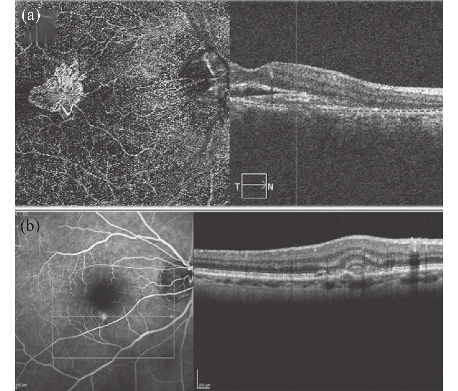
DE CARLO T E, ROMANO A, WAHEED N K, et al. A review of optical coherence tomography angiography (OCTA)[J]. International Journal of Retina and Vitreous, 2015, 1(1): 5. doi: 10.1186/s40942-015-0005-8
|
|
ZHANG Q Q, HUANG Y P, ZHANG T, et al. Wide-field imaging of retinal vasculature using optical coherence tomography-based microangiography provided by motion tracking[J]. Journal of Biomedical Optics, 2015, 20(6): 066008. doi: 10.1117/1.JBO.20.6.066008
|
|
WANG R K, AN L, FRANCIS P, et al. Depth-resolved imaging of capillary networks in retina and choroid using ultrahigh sensitive optical microangiography[J]. Optics Letters, 2010, 35(9): 1467-1469. doi: 10.1364/OL.35.001467
|
|
LI P, AN L, REIF R, et al. In vivo microstructural and microvascular imaging of the human corneo-scleral limbus using optical coherence tomography[J]. Biomedical Optics Express, 2011, 2(11): 3109-3118. doi: 10.1364/BOE.2.003109
|
|
ENFIELD J, JONATHAN E, LEAHY M. In vivo imaging of the microcirculation of the volar forearm using correlation mapping optical coherence tomography (cmOCT)[J]. Biomedical Optics Express, 2011, 2(5): 1184-1193. doi: 10.1364/BOE.2.001184
|
|
VAKOC B J, LANNING R M, TYRRELL J A, et al. Three-dimensional microscopy of the tumor microenvironment in vivo using optical frequency domain imaging[J]. Nature Medicine, 2009, 15(10): 1219-1223. doi: 10.1038/nm.1971
|
|
TANNO N, KISHI S. Optical coherence tomographic imaging and clinical diagnosis[J]. Medical Imaging Technology, 1999, 17: 3-10.
|
|
SCHUMAN J S, PULIAFITO C A, FUJIMOTO J G, et al.. Optical Coherence Tomography of Ocular Diseases[M]. 2nd ed. Thorofare, NJ: SLACK, New Jersey, 2004.
|
|
FANG L Y, CUNEFARE D, WANG CH, et al. Automatic segmentation of nine retinal layer boundaries in oct images of non-exudative amd patients using deep learning and graph search[J]. Biomedical Optics Express, 2017, 8(5): 2732-2744. doi: 10.1364/BOE.8.002732
|
|
DE OLIVEIRA DIAS J R, ZHANG Q Q, GARCIA J M B, et al. Natural history of subclinical neovascularization in nonexudative age-related macular degeneration using swept-source oct angiography[J]. Ophthalmology, 2018, 125(2): 255-266. doi: 10.1016/j.ophtha.2017.08.030
|
|
BOUTELEUX V, KODJIKIAN L, MENDES M, et al. Increased choroidal thickness: a new feature to monitor age-related macular degeneration recurrence[J]. Graefe’s Archive for Clinical and Experimental Ophthalmology, 2019, 257(4): 699-707. doi: 10.1007/s00417-018-04216-8
|
|
WU J H, SEBASTIAN R T, CHU C J, et al. Reduced macular vessel density and capillary perfusion in glaucoma detected using OCT angiography[J]. Current Eye Research, 2019, 44(5): 533-540. doi: 10.1080/02713683.2018.1563195
|
|
WANG J, HORMEL T T, YOU Q SH, et al. Robust non-perfusion area detection in three retinal plexuses using convolutional neural network in OCT angiography[J]. Biomedical Optics Express, 2020, 11(1): 330-345. doi: 10.1364/BOE.11.000330
|
|
CHAN V T T, SUN Z H, TANG SH M, et al. Spectral-domain OCT measurements in Alzheimer’s disease: a systematic review and meta-analysis[J]. Ophthalmology, 2019, 126(4): 497-510. doi: 10.1016/j.ophtha.2018.08.009
|
|
KAISER P K, BLODI B A, SHAPIRO H, et al. Angiographic and optical coherence tomographic results of the MARINA study of ranibizumab in neovascular age-related macular degeneration[J]. Ophthalmology, 2007, 114(10): 1868-1875. doi: 10.1016/j.ophtha.2007.04.030
|
|
APOSTOLOPOULOS M N, KOUTSANDREA C N, MOSCHOS M N, et al. Evaluation of successful macular hole surgery by optical coherence tomography and multifocal electroretinography[J]. American Journal of Ophthalmology, 2002, 134(5): 667-674. doi: 10.1016/S0002-9394(02)01700-2
|
|
TAN O, LI G, LU A T H, et al. Mapping of macular substructures with optical coherence tomography for glaucoma diagnosis[J]. Ophthalmology, 2008, 115(6): 949-956. doi: 10.1016/j.ophtha.2007.08.011
|
|
FUJIMOTO J, SWANSON E. The development, commercialization, and impact of optical coherence tomography[J]. Investigative Ophthalmology &Visual Science, 2016, 57(9): OCT1-OCT13.
|
|
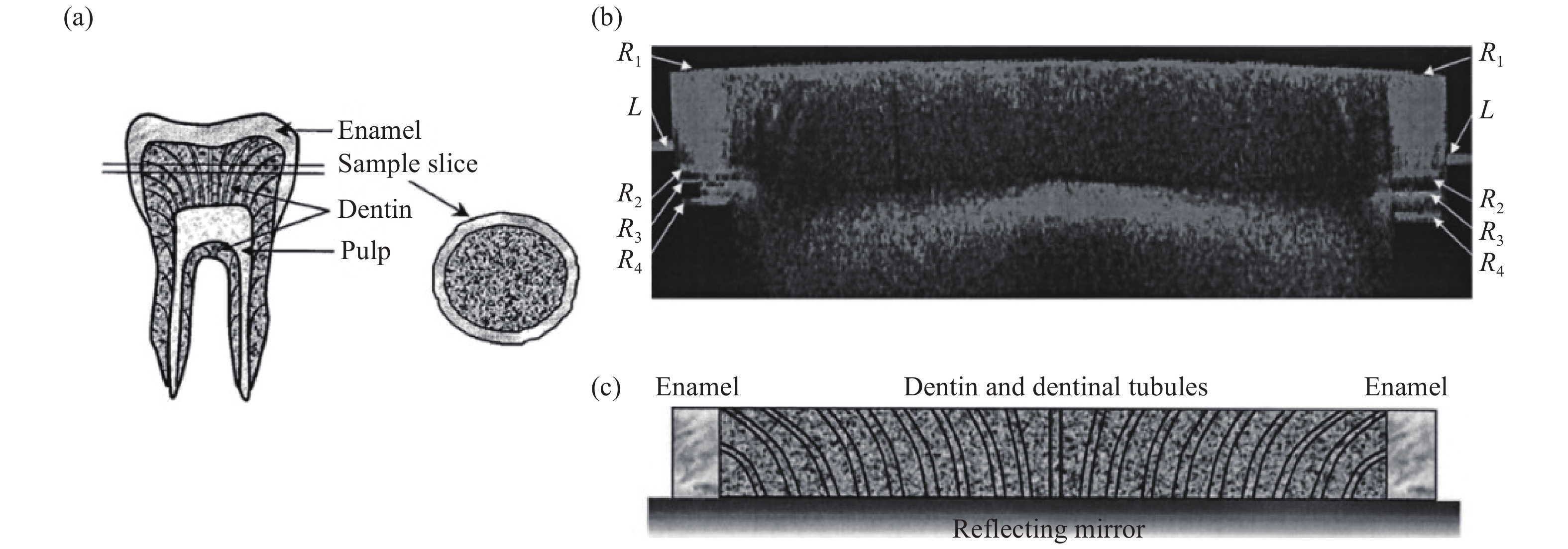
COLSTON B W, SATHYAM U S, DASILVA L B, et al. Dental OCT[J]. Optics Express, 1998, 3(6): 230-238. doi: 10.1364/OE.3.000230
|
|
WANG X J, MILNER T E, DE BOER J F, et al. Characterization of dentin and enamel by use of optical coherence tomography[J]. Applied Optics, 1999, 38(10): 2092-2096. doi: 10.1364/AO.38.002092
|
|
ADEN A, ANDERSON P, BURNETT G R, et al. Longitudinal correlation of 3D OCT to detect early stage erosion in bovine enamel[J]. Biomedical Optics Express, 2017, 8(2): 954-973. doi: 10.1364/BOE.8.000954
|
|
ZHOU Y, SHIMADA Y, MATIN K, et al. Assessment of root caries under wet and dry conditions using swept-source optical coherence tomography (SS-OCT)[J]. Dental Materials Journal, 2018, 37(6): 880-888. doi: 10.4012/dmj.2017-273
|
|
ALGHILAN M A, LIPPERT F, PLATT J A, et al. Impact of surface micromorphology and demineralization severity on enamel loss measurements by cross-polarization optical coherence tomography[J]. Journal of Dentistry, 2019, 81: 52-58. doi: 10.1016/j.jdent.2018.12.009
|
|
KUMAR ARAVETI S, HIRAISHI N, KOMINAMI N, et al. Swept-source optical coherence tomographic observation on prevalence and variations of cemento-enamel junction morphology[J]. Lasers in Medical Science, 2020, 35(1): 213-219. doi: 10.1007/s10103-019-02847-9
|
|
GAMBICHLER T, JAEDICKE V, TERRAS S. Optical coherence tomography in dermatology: technical and clinical aspects[J]. Archives of Dermatological Research, 2011, 303(7): 457-473. doi: 10.1007/s00403-011-1152-x
|
|
SATTLER E C, KÄSTLE R, WELZEL J. Optical coherence tomography in dermatology[J]. Journal of Biomedical Optics, 2013, 18(6): 061224. doi: 10.1117/1.JBO.18.6.061224
|
|
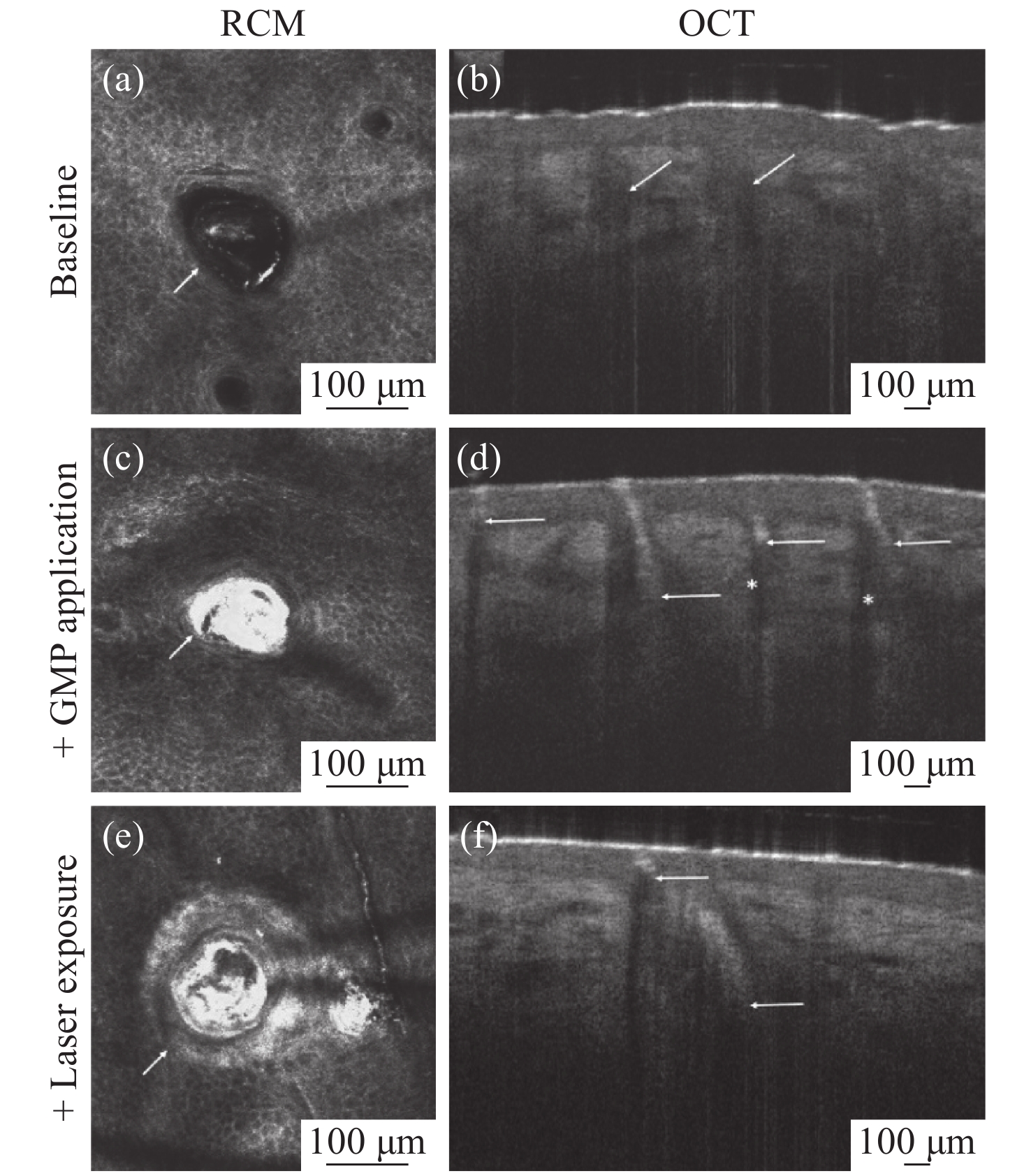
FUCHS C S K, ORTNER V K, MOGENSEN M, et al. Transfollicular delivery of gold microparticles in healthy skin and acne vulgaris, assessed by in vivo reflectance confocal microscopy and optical coherence tomography[J]. Lasers in Surgery and Medicine, 2019, 51(5): 430-438. doi: 10.1002/lsm.23076
|
|
MOGENSEN M, THRANE L, JØRGENSEN T M, et al. OCT imaging of skin cancer and other dermatological diseases[J]. Journal of Biophotonics, 2009, 2(6-7): 442-451. doi: 10.1002/jbio.200910020
|
|
MOGENSEN M, NÜRNBERG B M, FORMAN J L, et al. In vivo thickness measurement of basal cell carcinoma and actinic keratosis with optical coherence tomography and 20-MHz ultrasound[J]. British Journal of Dermatology, 2009, 160(5): 1026-1033. doi: 10.1111/j.1365-2133.2008.09003.x
|
|
KRATKIEWICZ K, MANWAR R, RAJABI-ESTARABADI A, et al. Photoacoustic/ultrasound/optical coherence tomography evaluation of melanoma lesion and healthy skin in a swine model[J]. Sensors, 2019, 19(12): 2815. doi: 10.3390/s19122815
|
|
ZHAO Y, CHU K K, WAX A. Enhanced depth penetration by dual-axis optical coherence tomography[J]. Proceedings of SPIE, 2019, 10867: 1086704.
|
|
YOW A P, SRIVASTAVA R, CHENG J, et al.. Techniques and Applications in Skin OCT Analysis[M]. Springer, 2020.
|
|
AKASAKA T, KUBO T, MIZUKOSHI M, et al. Pathophysiology of acute coronary syndrome assessed by optical coherence tomography[J]. Journal of Cardiology, 2010, 56(1): 8-14. doi: 10.1016/j.jjcc.2010.05.005
|
|
COSTOPOULOS C, BROWN A J, TENG ZH ZH, et al. Intravascular ultrasound and optical coherence tomography imaging of coronary atherosclerosis[J]. International Journal of Cardiovascular Imaging, 2016, 32(1): 189-200. doi: 10.1007/s10554-015-0701-3
|
|
SUN R, SUN L P, FU Y D, et al. Culprit plaque characteristics in women vs men with a first ST-segment elevation myocardial infarction: in vivo optical coherence tomography insights[J]. Clinical Cardiology, 2017, 40(12): 1285-1290. doi: 10.1002/clc.22825
|
|
LIANG Y T, LIU L W, HU S Y, et al. Characterizing physical properties and in vivo OCT imaging study of Cu-Sn-S nanocrystals[J]. AIP Advances, 2017, 7(1): 015012. doi: 10.1063/1.4973731
|
|
YANG SH ZH, CHEN H B, LIU L W, et al. OCT imaging detection of brain blood vessels in mouse, based on semiconducting polymer nanoparticles[J]. Analyst, 2017, 142(23): 4503-4510. doi: 10.1039/C7AN01245D
|
|
LIU L W, HU S Y, WANG Y, et al. Optimizing the synthesis of core/shell structure Au@Cu2S nanocrystals as contrast-enhanced for bioimaging detection[J]. Scientific Reports, 2018, 8(1): 8866. doi: 10.1038/s41598-018-27015-x
|
|
MURATA A, WALLACE-BRADLEY D, TELLEZ A, et al. Accuracy of optical coherence tomography in the evaluation of neointimal coverage after stent implantation[J]. JACC:Cardiovascular Imaging, 2010, 3(1): 76-84. doi: 10.1016/j.jcmg.2009.09.018
|
|
PERKINS L E L, RIPPY M K. Balloons and stents and scaffolds: preclinical evaluation of interventional devices for occlusive arterial disease[J]. Toxicologic Pathology, 2019, 47(3): 297-310. doi: 10.1177/0192623318815604
|
|
LIU X L, SUN CH B, TIAN J T, et al. Shrinkage as a potential mechanism of recurrent events in patients with a large vulnerable plaque[J]. Journal of Cardiovascular Medicine, 2019, 20(8): 518-524. doi: 10.2459/JCM.0000000000000783
|
|
WANG J Q, PARITALA P K, MENDIETA J B, et al. Optical coherence tomography-based patient-specific coronary artery reconstruction and fluid–structure interaction simulation[J]. Biomechanics and Modeling in Mechanobiology, 2020, 19(1): 7-20. doi: 10.1007/s10237-019-01191-9
|
|
NAKAMURA T, HORIKOSHI T, KUGIYAMA K. Relationship of a thinned medial layer to the attenuated contractile response in atherosclerotic coronary arteries[J]. American Journal of Physiology-Heart and Circulatory Physiology, 2020, 318(1): H135-H142. doi: 10.1152/ajpheart.00537.2019
|
|
KHOLODNYKH A I, PETROVA I Y, LARIN K V, et al. Precision of measurement of tissue optical properties with optical coherence tomography[J]. Applied Optics, 2003, 42(16): 3027-3037. doi: 10.1364/AO.42.003027
|
|
SONG Y CH, GARCIA S, FROMETA Y, et al. Quantitative assessment of hemodynamic and structural characteristics of in vivo brain tissue using total diffuse reflectance spectrum measured in a non-contact fashion[J]. Biomedical Optics Express, 2017, 8(1): 78-103. doi: 10.1364/BOE.8.000078
|
|
SU Y, YAO X S, LI ZH H, et al. Measurements of the thermal coefficient of optical attenuation at different depth regions of in vivo human skins using optical coherence tomography: a pilot study[J]. Biomedical Optics Express, 2015, 6(2): 500-513. doi: 10.1364/BOE.6.000500
|
|
MEI L, SOMESFALEAN G, SVANBERG S. Frequency-modulated light scattering interferometry employed for optical properties and dynamics studies of turbid media[J]. Biomedical Optics Express, 2014, 5(8): 2810-2822. doi: 10.1364/BOE.5.002810
|
|
KHOLODNYKH A I, PETROVA I Y, MOTAMEDI M, et al. Accurate measurement of total attenuation coefficient of thin tissue with optical coherence tomography[J]. IEEE Journal of Selected Topics in Quantum Electronics, 2003, 9(2): 210-221. doi: 10.1109/JSTQE.2003.814194
|
|
LI K Y, LIANG W X, YANG Z H, et al. Robust, accurate depth-resolved attenuation characterization in optical coherence tomography[J]. Biomedical Optics Express, 2020, 11(2): 672-687. doi: 10.1364/BOE.382493
|
|
LEVITZ D, THRANE L, FROSZ M H, et al. Determination of optical scattering properties of highly-scattering media in optical coherence tomography images[J]. Optics Express, 2004, 12(2): 249-259. doi: 10.1364/OPEX.12.000249
|
|
SORIN W V, GRAY D F. Simultaneous thickness and group index measurement using optical low-coherence reflectometry[J]. IEEE Photonics Technology Letters, 1992, 4(1): 105-107. doi: 10.1109/68.124892
|
|
WANG X J, MILNER T E, DHOND R P, et al. Characterization of human scalp hairs by optical low-coherence reflectometry[J]. Optics Letters, 1995, 20(6): 524-526. doi: 10.1364/OL.20.000524
|
|
WANG X J, MILNER T E, CHANG M C, et al. Group refractive index measurement of dry and hydrated type I collagen films using optical low-coherence reflectometry[J]. Journal of Biomedical Optics, 1996, 1(2): 212-216. doi: 10.1117/12.227699
|
|
ZVYAGIN A V, SILVA K K M B D, ALEXANDROV S A, et al. Refractive index tomography of turbid media by bifocal optical coherence refractometry[J]. Optics Express, 2003, 11(25): 3503-3517. doi: 10.1364/OE.11.003503
|
Figures(8)


 DownLoad: