


-
摘要: 多光子成像技术是一种层析能力好、信噪比高的新型光学成像技术。在皮肤光学三维检测中,多光子技术已经应用于无创在体成像,且已得到产业化开发。本文将首先介绍多光子皮肤检测系统的若干核心技术,即双光子自发荧光技术、二次谐波成像技术、荧光寿命成像技术、相干反斯托克斯-拉曼成像技术等,然后简要介绍多光子成像系统在皮肤疾病成像检测上的应用,最后分析该系统的优势和未来可能的发展趋势。
-
关键词:
- 皮肤组织成像 /
- 多光子荧光成像 /
- 荧光寿命成像 /
- 相干反斯托克斯-拉曼 /
- 皮肤衰老检测
Abstract: Multi-photon imaging is a new technique for optical imaging with inherent optical sectioning abilities and a high signal-to-noise ratio. Multi-photon microscopy has applications in dermatological imaging for its non-invasive and in situ imaging capability. As such, it has been applied commercially. In this paper, we first introduce the core technologies of multiphoton microscopy including two-photon auto-fluorescence imaging, second-harmonic generation(SHG), fluorescence life time imaging(FLIM) and coherent anti-Stokes Raman scattering(CARS). We then introduce its application on skin imaging analysis. Finally, we analyze the advantages and the possible future development of this technology. -


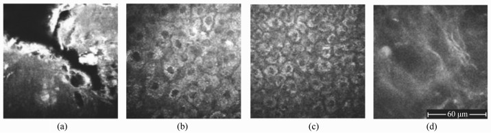
图 2 健康人体皮肤活体成像,激发光波长760 nm:(a)角质层,0 μm深:角蛋白自发荧光信号;(b)颗粒层,20 μm深:角质细胞中的透明角质颗粒、NADPH、角蛋白自发荧光信号;(c)棘层,30 μm深:角质细胞密度增大;激发光波长800 nm; (d)真皮层,85 μm深:胶原蛋白、弹性蛋白自发荧光信号[11]
Figure 2. In vivo healthy human skin imaging with excitation wavelength of 760 nm:(a)Stratum corneum, 0 μm depth: auto-fluorescence signal of keratin; (b)Stratum granulosum, 20 μm depth: auto-fluorescence signal of keratohyalin granules, NADPH, and keratin in keratinocytes; (c)Stratum spinosum, 30 μm depth: increased cellular density of keratinocytes; 800 nm excitation wavelength: (d)Dermis, 85 μm depth: auto-fluorescence signal of collagen and elastin[11]


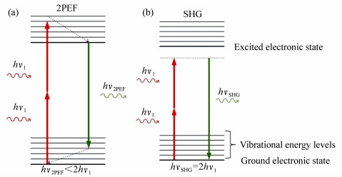
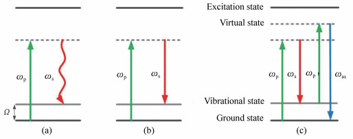
图 6 相干反斯托克斯-拉曼(CARS)的Jablonski图:(a)拉曼散射; (b)受激拉曼散射; (c)相干反斯托克斯-拉曼
Figure 6. Jablonski image of coherent anti-Stokes Raman scattering(CARS):(a)Raman Scattering; (b)Stimulated Raman Scattering; (c)Coherent Anti-Stokes Raman Scattering

-
[1] KONIG K, RIEMANN I. High-resolution multiphoton tomography of human skin with subcellular spatial resolution and picosecond time resolution[J]. J. Biomed Opt., 2003, 8(3):432-439. doi: 10.1117/1.1577349 [2] ZHANG Y, HONG H, CAI W. Photoacoustic imaging[J]. Cold Spring Harb Protoc, 2011, 2011(9):1015-1025. http://d.old.wanfangdata.com.cn/Periodical/gzxb200507015 [3] HUANG D, SWANSON E A, LIN C P, et al.. Optical coherence tomography[J]. Science, 1991, 254(5035):1178-1181. doi: 10.1126/science.1957169 [4] DENK W, STRICKLER J H, WEBB W W. Two-photon laser scanning fluorescence microscopy[J]. Science, 1990, 248(4951):73-76. doi: 10.1126/science.2321027 [5] KÖNIG K. Multiphoton Tomography[OL].[2018-02-20].http:www.mpt-tomography.de. [6] KÖNIG K. Clinical multiphoton tomography[J]. J. Biophotonics, 2008, 1(1):13-23. doi: 10.1002/(ISSN)1864-0648 [7] BREUNIG H, WEINIGEL M, BVCKLE R, et al.. Clinical coherent anti-Stokes Raman scattering and multiphoton tomography of human skin with a femtosecond laser and photonic crystal fiber[J]. Laser Physics Letters, 2013, 10(2):025604. doi: 10.1088/1612-2011/10/2/025604 [8] MONICI M. Cell and tissue autofluorescence research and diagnostic applications[J]. Biotechnol Annu Rev, 2005, 11:227-256. doi: 10.1016/S1387-2656(05)11007-2 [9] 席鹏, 刘宇嘉, 姚志荣, 等.用于皮肤影像诊断的光学成像方法[J].中国激光, 2011, 38(2):7-19. http://www.wanfangdata.com.cn/details/detail.do?_type=perio&id=QK201100094508XI P, LIU Y J, YAO ZH R, et al.. Optical imaging techniques in skin imaging diagnosis[J]. Chinese Journal of Lasers, 2011, 38(2):7-19.(in Chinese) http://www.wanfangdata.com.cn/details/detail.do?_type=perio&id=QK201100094508 [10] JAMME F, KASCAKOVA S, VILLETTE S, et al.. Deep UV autofluorescence microscopy for cell biology and tissue histology[J]. Biol. Cell, 2013, 105(7):277-288. doi: 10.1111/boc.201200075 [11] SEIDENARI S, ARGINELLI F, BASSOLI S, et al.. Multiphoton laser microscopy and fluorescence lifetime imaging for the evaluation of the skin[J]. Dermatol Res Pract, 2012, 2012:810749. http://d.old.wanfangdata.com.cn/OAPaper/oai_doaj-articles_68181436a763faeb55a9616ae70c9214 [12] GIBSON E A, MASIHZADEH O, LEI T C, et al.. Multiphoton microscopy for ophthalmic imaging[J]. Journal of Ophthalmology, 2011, 2011:870879. http://d.old.wanfangdata.com.cn/OAPaper/oai_pubmedcentral.nih.gov_3022205 [13] BALU M, ZACHARY C B, HARRIS R M, et al.. In Vivo multiphoton microscopy of basal cell carcinoma[J]. JAMA Dermatol, 2015, 151(10):1068-1074. doi: 10.1001/jamadermatol.2015.0453 [14] TSAI T H, JEE S H, DONG C Y, et al.. Multiphoton microscopy in dermatological imaging[J]. J. Dermatol Sci., 2009, 56(1):1-8. http://d.old.wanfangdata.com.cn/OAPaper/oai_pubmedcentral.nih.gov_3233249 [15] KÖNIG K. Fluorescence Lifetime Imaging[OL].[2018-02-20].http://mpt-tomography.de/fluorescence-lifetime-imaging. [16] DANCIK Y, FAVRE A, LOY C.J, et al.. Use of multiphoton tomography and fluorescence lifetime imaging to investigate skin pigmentation in vivo[J]. J. Biomed Opt., 2013, 18(2):26022. doi: 10.1117/1.JBO.18.2.026022 [17] WEINIGEL M, BREUNIG H G, KELLNER-HÖFER M, et al.. In vivo histology:optical biopsies with chemical contrast using clinical multiphoton/coherent anti-Stokes Raman scattering tomography[J]. Laser Physics Letters, 2014, 11(5):055601. doi: 10.1088/1612-2011/11/5/055601 [18] KÖNIG K. In-Vivo Clinical Applications[OL].[2018-02-20]. http://mpt-tomography.de/vivo-clinical-applications. [19] KOEHLER M J, HAHN S, PRELLER A, et al.. Morphological skin ageing criteria by multiphoton laser scanning tomography:non-invasive in vivo scoring of the dermal fibre network[J]. Exp. Dermatol, 2008, 17(6):519-523. doi: 10.1111/j.1600-0625.2007.00669.x [20] LIN S J, WU R, J R, TAN H Y, et al.. Evaluating cutaneous photoaging by use of multiphoton fluorescence and second-harmonic generation microscopy[J]. Opt. Lett., 2005, 30(17):2275-2277. doi: 10.1364/OL.30.002275 [21] SONG W, XU Q, ZHANG Y, et al.. Fully integrated reflection-mode photoacoustic, two-photon, and second harmonic generation microscopy in vivo[J]. Sci. Rep., 2016, 6:32240. doi: 10.1038/srep32240 -

 下载:
下载:


计量
- 文章访问数: 4432
- HTML全文浏览量: 1740
- PDF下载量: 330
- 被引次数: 0
